Park Byeong-Ung, Park Sang Min, Lee Kyoung-Pil, Lee Seong Jin, Nam Yu Eun, Park Han Sang, Eom Seongsu, Lim Jeong Ok, Kim Dong Sung, Kim Hong Kyun
Bio-Medical Institute, Kyungpook National University Hospital (KNUH), Daegu, South Korea.
Department of Ophthalmology, School of Medicine, Kyungpook National University, Daegu, South Korea.
J Tissue Eng. 2019 Nov 14;10:2041731419887833. doi: 10.1177/2041731419887833. eCollection 2019 Jan-Dec.
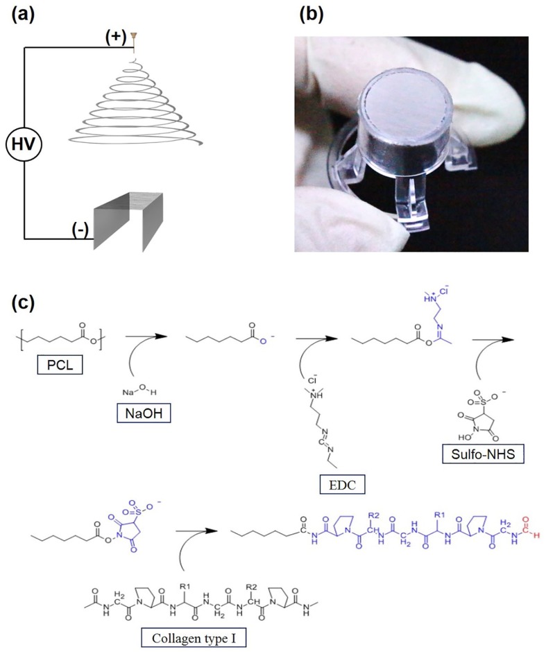
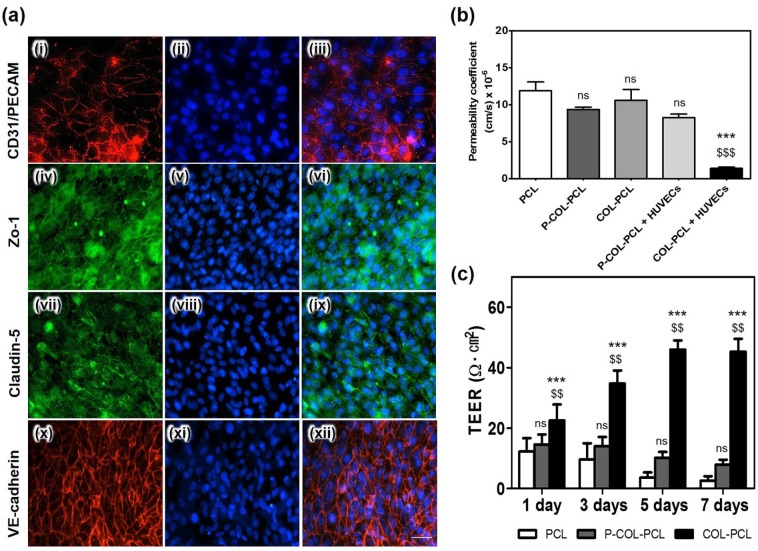
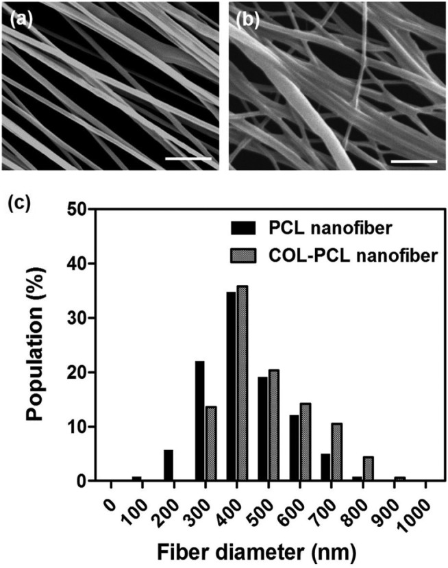
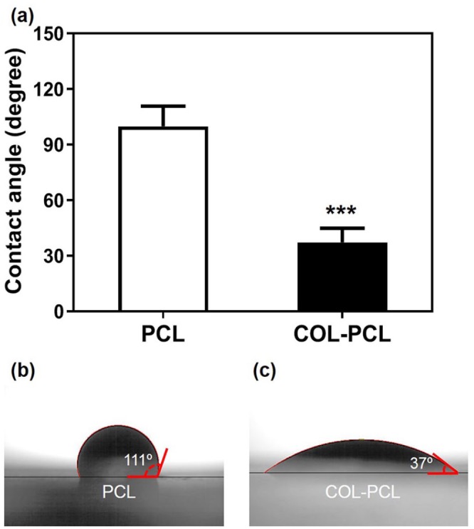
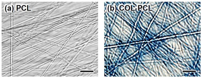
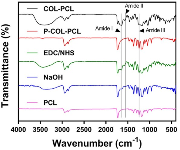
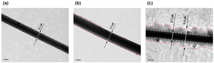
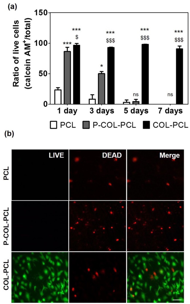
The endothelialization on the poly (ε-caprolactone) nanofiber has been limited due to its low hydrophilicity. The aim of this study was to immobilize collagen on an ultra-thin poly (ε-caprolactone) nanofiber membrane without altering the nanofiber structure and maintaining the endothelial cell homeostasis on it. We immobilized collagen on the poly (ε-caprolactone) nanofiber using hydrolysis by NaOH treatment and 1-ethyl-3-(3-dimethylaminopropyl) carbodiimide/sulfo--hydroxysulfosuccinimide reaction as a cost-effective and stable approach. NaOH was first applied to render the poly (ε-caprolactone) nanofiber hydrophilic. Subsequently, collagen was immobilized on the surface of the poly (ε-caprolactone) nanofibers using 1-ethyl-3-(3-dimethylaminopropyl) carbodiimide/sulfo--hydroxysulfosuccinimide. Scanning electron microscopy, Fourier transform infrared spectroscopy, transmission electron microscopy, and fluorescence microscopy were used to verify stable collagen immobilization on the surface of the poly (ε-caprolactone) nanofibers and the maintenance of the original structure of poly (ε-caprolactone) nanofibers. Furthermore, human endothelial cells were cultured on the collagen-immobilized poly (ε-caprolactone) nanofiber membrane and expressed tight junction proteins with the increase in transendothelial electrical resistance, which demonstrated the maintenance of the endothelial cell homeostasis on the collagen-immobilized-poly (ε-caprolactone) nanofiber membrane. Thus, we expected that this process would be promising for maintaining cell homeostasis on the ultra-thin poly (ε-caprolactone) nanofiber scaffolds.
聚(ε-己内酯)纳米纤维的内皮化由于其低亲水性而受到限制。本研究的目的是在不改变纳米纤维结构的情况下,将胶原蛋白固定在超薄聚(ε-己内酯)纳米纤维膜上,并维持其上的内皮细胞稳态。我们使用氢氧化钠处理水解和1-乙基-3-(3-二甲基氨基丙基)碳二亚胺/磺基-羟基琥珀酰亚胺反应,作为一种经济高效且稳定的方法,将胶原蛋白固定在聚(ε-己内酯)纳米纤维上。首先应用氢氧化钠使聚(ε-己内酯)纳米纤维具有亲水性。随后,使用1-乙基-3-(3-二甲基氨基丙基)碳二亚胺/磺基-羟基琥珀酰亚胺将胶原蛋白固定在聚(ε-己内酯)纳米纤维表面。通过扫描电子显微镜、傅里叶变换红外光谱、透射电子显微镜和荧光显微镜来验证胶原蛋白在聚(ε-己内酯)纳米纤维表面的稳定固定以及聚(ε-己内酯)纳米纤维原始结构的维持。此外,将人内皮细胞培养在固定有胶原蛋白的聚(ε-己内酯)纳米纤维膜上,随着跨内皮电阻的增加,细胞表达紧密连接蛋白,这表明在固定有胶原蛋白的聚(ε-己内酯)纳米纤维膜上维持了内皮细胞稳态。因此,我们期望这一过程对于在超薄聚(ε-己内酯)纳米纤维支架上维持细胞稳态具有广阔前景。