Center for Biomechanics and Bioengineering, Key Laboratory of Microgravity (National Microgravity Laboratory) and Beijing Key Laboratory of Engineered Construction and Mechanobiology, Institute of Mechanics, Chinese Academy of Sciences, Beijing, 100190, China.
School of Engineering Science, University of Chinese Academy of Sciences, Beijing, China.
Stem Cell Res Ther. 2019 Nov 27;10(1):349. doi: 10.1186/s13287-019-1454-z.
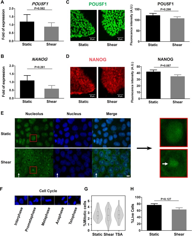
Distinct mechanical stimuli are known to manipulate the behaviors of embryonic stem cells (ESCs). Fundamental rationale of how ESCs respond to mechanical forces and the potential biological effects remain elusive. Here we conducted the mechanobiological study for hESCs upon mechanomics analysis to unravel typical mechanosensitive processes on hESC-specific fluid shear.
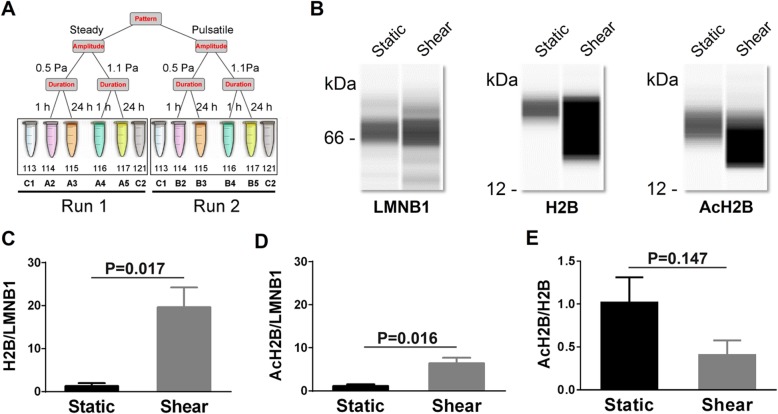
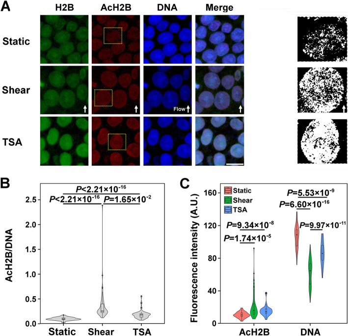
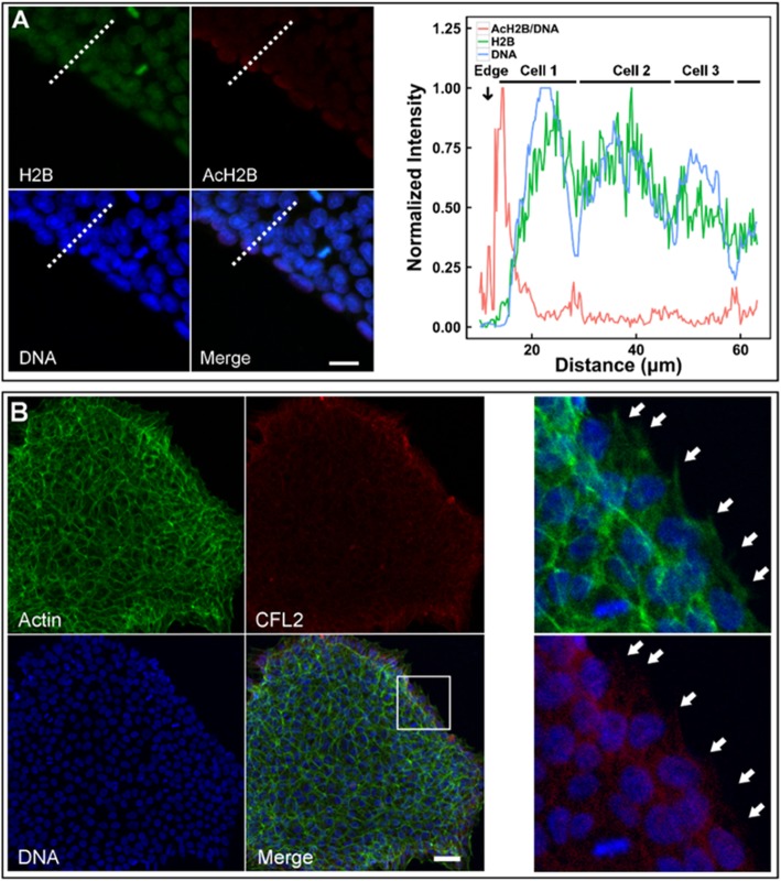
hESC line H1 was subjected to systematically varied shear flow, and mechanosensitive proteins were obtained by mass spectrometry (MS) analysis. Then, function enrichment analysis was performed to identify the enriched gene sets. Under a steady shear flow of 1.1 Pa for 24 h, protein expressions were further detected using western blotting (WB), quantitative real-time PCR (qPCR), and immunofluorescence (IF) staining. Meanwhile, the cells were treated with 200 nM trichostatin (TSA) for 1 h as positive control to test chromatin decondensation. Actin, DNA, and RNA were then visualized with TRITC-labeled phalloidin, Hoechst 33342, and SYTO® RNASelect™ green fluorescent cell stain (Life Technologies), respectively. In addition, cell stiffness was determined with atomic force microscopy (AFM) and annexin V-PE was used to determine the apoptosis with a flow cytometer (FCM).
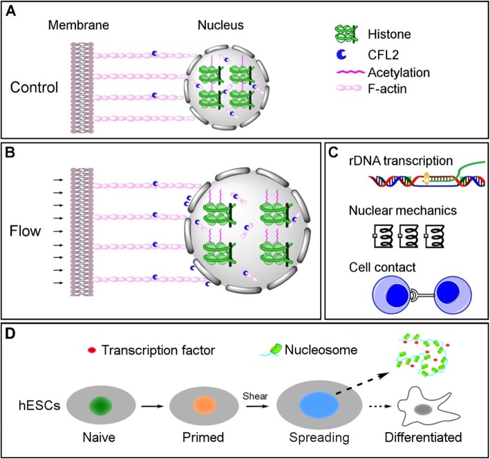
Typical mechanosensitive proteins were unraveled upon mechanomics analysis under fluid shear related to hESCs in vivo. Functional analyses revealed significant alterations in histone acetylation, nuclear size, and cytoskeleton for hESC under shear flow. Shear flow was able to induce H2B acetylation and nuclear spreading by CFL2/F-actin cytoskeletal reorganization. The resulting chromatin decondensation and a larger nucleus readily accommodate signaling molecules and transcription factors.
Shear flow regulated chromatin dynamics in hESCs via cytoskeleton and nucleus alterations and consolidated their primed state.
已知不同的机械刺激可以操纵胚胎干细胞(ESCs)的行为。ESCs 如何响应机械力以及潜在的生物学效应的基本原理仍然难以捉摸。在这里,我们通过机械组学分析对 hESC 进行了力学生物学研究,以揭示 hESC 特异性流体剪切力下的典型力学敏感过程。
将 hESC 系 H1 置于系统变化的剪切流中,通过质谱(MS)分析获得机械敏感蛋白。然后,进行功能富集分析以识别富集的基因集。在 1.1 Pa 的稳态剪切流中孵育 24 小时后,使用 Western blot(WB)、定量实时 PCR(qPCR)和免疫荧光(IF)染色进一步检测蛋白质表达。同时,将细胞用 200 nM 曲古抑菌素(TSA)处理 1 小时作为阳性对照,以测试染色质解凝聚。然后,用 TRITC 标记的鬼笔环肽、Hoechst 33342 和 SYTO®RNASelect™绿色荧光细胞染色剂(Life Technologies)分别可视化肌动蛋白、DNA 和 RNA。此外,通过原子力显微镜(AFM)测定细胞刚度,并使用 Annexin V-PE 通过流式细胞仪(FCM)测定细胞凋亡。
在与体内 hESC 相关的流体剪切下的机械组学分析中揭示了典型的机械敏感蛋白。功能分析显示,剪切流下 hESC 的组蛋白乙酰化、核大小和细胞骨架发生显著变化。剪切流能够通过 CFL2/F-肌动蛋白细胞骨架重排诱导 H2B 乙酰化和核铺展。由此产生的染色质解凝聚和更大的核更容易容纳信号分子和转录因子。
剪切流通过细胞骨架和核的改变调节 hESC 中的染色质动力学,并巩固其初始状态。