Atsumi Hideki, Horie Tomohiko, Kajihara Nao, Sunaga Azusa, Sakakibara Yumetaro, Matsumae Mitsunori
Department of Neurosurgery, Tokai University School of Medicine.
Division of Diagnostic Image Analysis, Tohoku University Graduate School of Medicine.
Neurol Med Chir (Tokyo). 2020 Jan 15;60(1):30-36. doi: 10.2176/nmc.oa.2019-0170. Epub 2019 Nov 27.
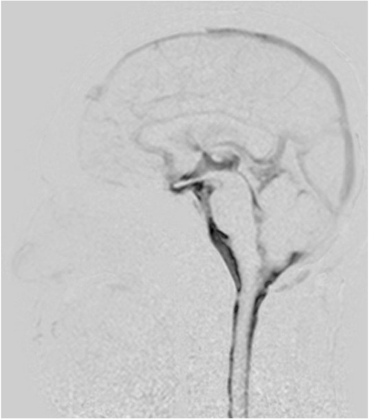
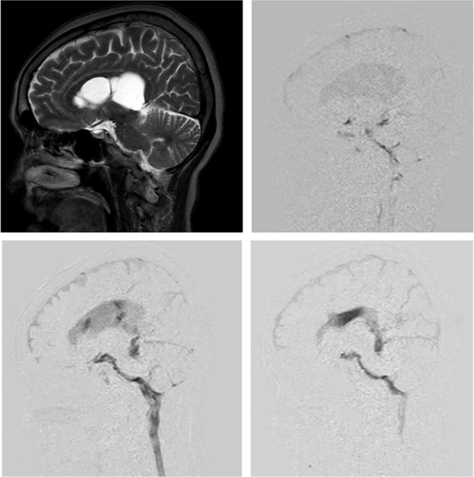
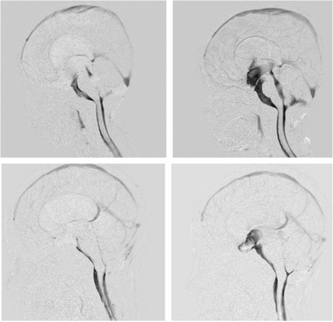
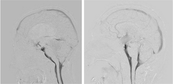
The motion of cerebrospinal fluid (CSF) within the subarachnoid space and ventricles is greatly modulated when propagating synchronously with the cardiac pulse and respiratory cycle and path through the nerves, blood vessels, and arachnoid trabeculae. Water molecule movement that propagates between two spaces via a stoma, foramen, or duct presents increased acceleration when passing through a narrow area and can exhibit "turbulence." Recently, neurosurgeons have started to perform fenestration procedures using neuroendoscopy to treat hydrocephalus and cystic lesions. As part of the postoperative evaluation, a noninvasive diagnostic technique to visualize the water molecules at the fenestrated site is necessary. Because turbulence is observed at this fenestrated site, an imaging technique appropriate for observing this turbulence is essential. We therefore investigated the usefulness of a dynamic improved motion-sensitized driven-equilibrium steady-state free precession (Dynamic iMSDE SSFP) sequence of magnetic resonance imaging that is superior for ascertaining turbulent motions in healthy volunteers and patients. Images of Dynamic iMSDE SSFP from volunteers revealed that CSF motion at the ventral surface of the brainstem and the third ventricle is augmented and turbulent. Moreover, our findings confirmed that this technique is useful for evaluating treatments that utilize neuroendoscopy. As a result, Dynamic iMSDE SSFP, a simple sequence for visualizing CSF motion, entails a short imaging time, can extensively visualize CSF motion, does not require additional processes such as labeling or trigger setting, and is anticipated to have wide-ranging clinical applications in the future.
当脑脊液(CSF)在蛛网膜下腔和脑室内的流动与心脏搏动和呼吸周期同步传播并通过神经、血管和蛛网膜小梁时,其运动受到极大调节。水分子通过小孔、孔道或导管在两个空间之间传播时,在通过狭窄区域时会出现加速度增加,并可能表现出“湍流”。最近,神经外科医生已开始使用神经内镜进行开窗手术来治疗脑积水和囊性病变。作为术后评估的一部分,需要一种非侵入性诊断技术来可视化开窗部位的水分子。由于在该开窗部位观察到湍流,因此适合观察这种湍流的成像技术至关重要。因此,我们研究了磁共振成像的动态改进运动敏感驱动平衡稳态自由进动(Dynamic iMSDE SSFP)序列在健康志愿者和患者中确定湍流运动方面的有用性。志愿者的Dynamic iMSDE SSFP图像显示,脑干腹侧表面和第三脑室的脑脊液运动增强且呈湍流状态。此外,我们的研究结果证实,该技术对于评估利用神经内镜的治疗方法很有用。因此,Dynamic iMSDE SSFP作为一种可视化脑脊液运动的简单序列,成像时间短,可以广泛地可视化脑脊液运动,不需要标记或触发设置等额外过程,预计未来将有广泛的临床应用。