Luther Evan, Matus Alejandro, Eichberg Daniel G, Shah Ashish H, Ivan Michael
Neurological Surgery, University of Miami Miller School of Medicine, Miami, USA.
Neurological Surgery, Florida International University, Herbert Wertheim College of Medicine, Miami, USA.
Cureus. 2019 Oct 14;11(10):e5905. doi: 10.7759/cureus.5905.
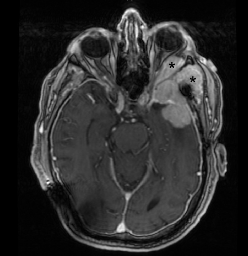
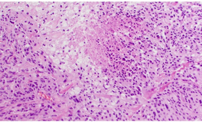
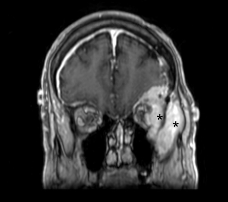
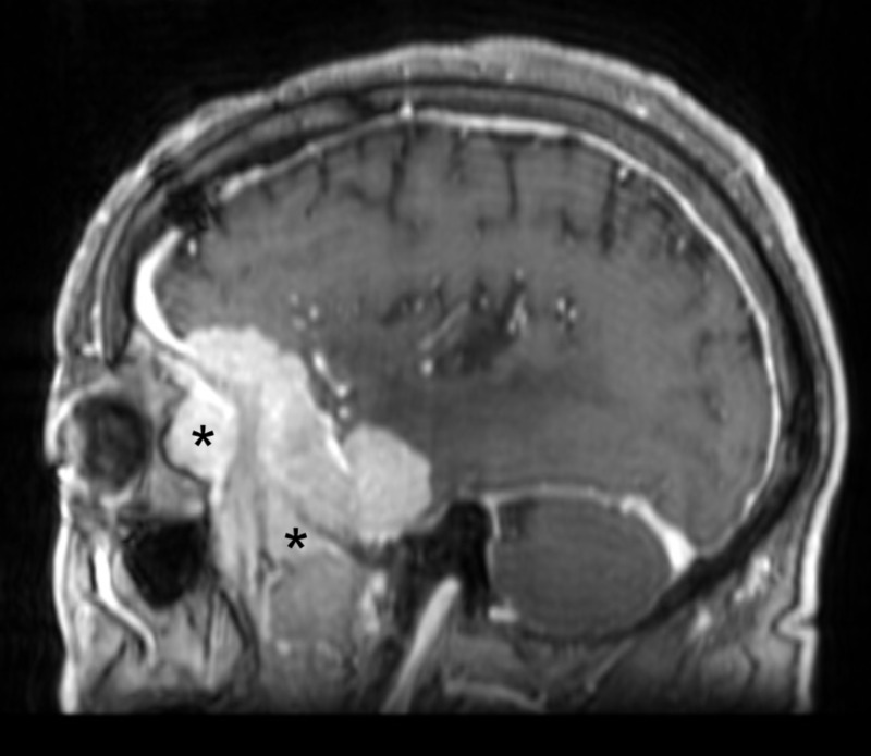
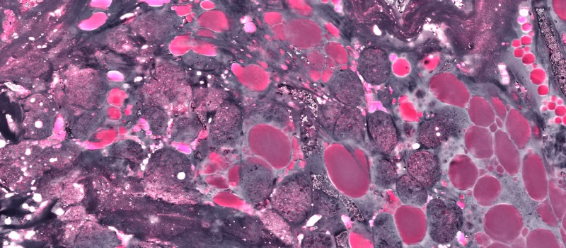
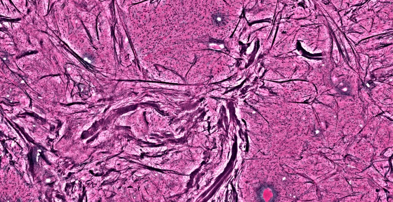
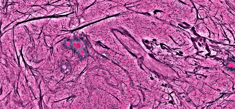
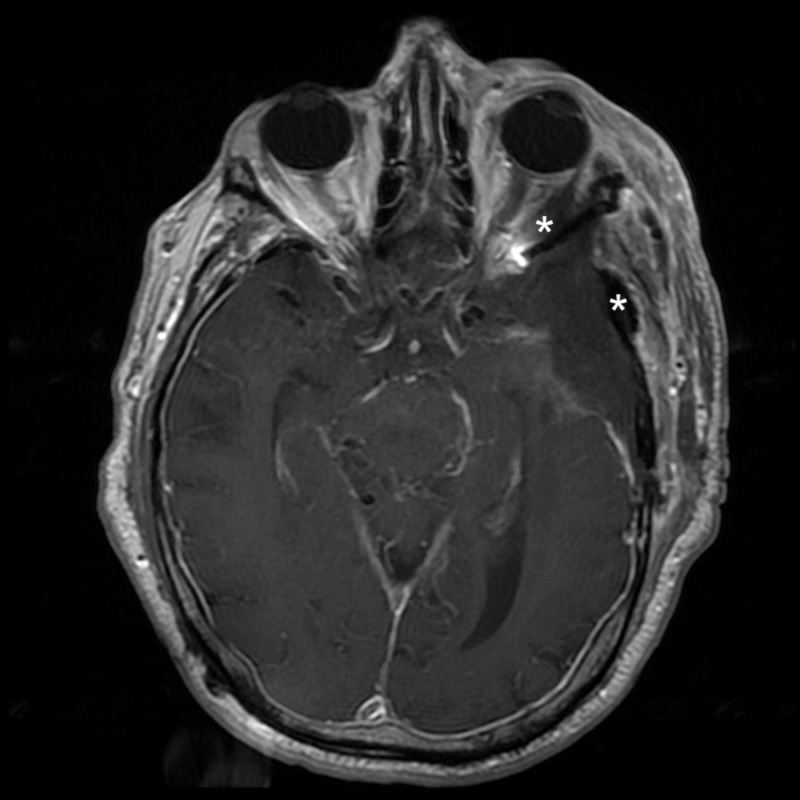
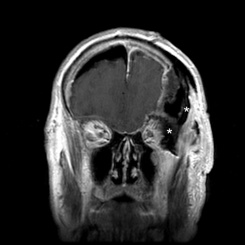
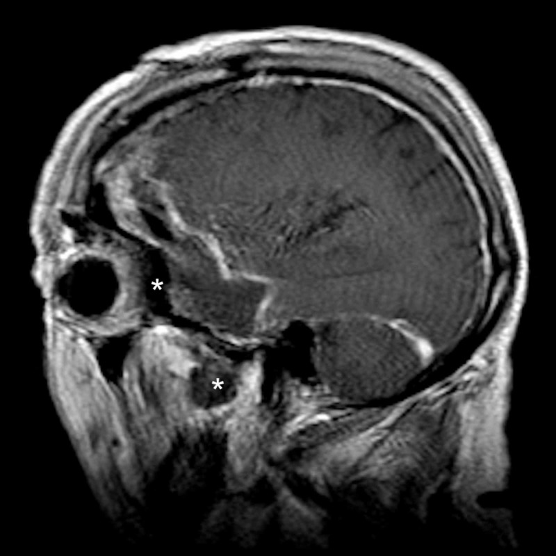
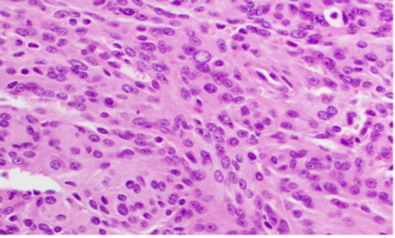
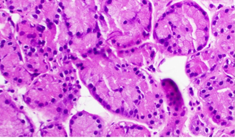
Meningiomas are the most common intracranial, extra-axial neoplasms and account for a significant proportion of all central nervous system (CNS) tumors. Regardless of the grade, treatment typically involves upfront surgical resection. However, in many instances, especially in meningiomas arising from the skull base, complete removal is often difficult given the close proximity to important anatomic structures. In this report, we discuss the use of stimulated Raman histology as a means to identify tissue boundaries during the resection of an extensive, recurrent, atypical spheno-orbital meningioma. We report a 75-year-old male with a history of a prior left frontotemporal craniotomy for a grade II meningioma three years prior, who presented with worsening left-sided visual loss and pronounced temporal bossing. Repeat magnetic resonance imaging (MRI) revealed a recurrent left spheno-orbital tumor suggestive of a meningioma extending into the middle cranial fossa, the lateral orbit, and the temporalis muscle. He underwent an extended orbito-pterional craniotomy, and intraoperative stimulated Raman histology aided in the identification of tumor margins within the orbit and the temporalis muscle in order to better preserve the normal orbital contents and muscle bulk of the infratemporal fossa. This case demonstrates the utility of stimulated Raman histology during the resection of invasive skull base tumors. The immediate intraoperative Raman histologic sections can clearly identify tissue boundaries and thus help preserve important anatomic structures. Continued development of this method can potentially improve the accuracy of intraoperative diagnoses and guide surgeons during tumor resections near eloquent anatomical regions or important normal structures.
脑膜瘤是最常见的颅内、轴外肿瘤,在所有中枢神经系统(CNS)肿瘤中占相当大的比例。无论分级如何,治疗通常首先进行手术切除。然而,在许多情况下,尤其是起源于颅底的脑膜瘤,由于与重要解剖结构距离很近,往往难以完全切除。在本报告中,我们讨论了使用受激拉曼组织学作为一种手段,在广泛的、复发性、非典型蝶眶脑膜瘤切除术中识别组织边界。我们报告了一名75岁男性,三年前曾因II级脑膜瘤接受左额颞开颅手术,此次因左侧视力恶化和明显的颞部隆起就诊。重复磁共振成像(MRI)显示左侧蝶眶肿瘤复发,提示脑膜瘤延伸至中颅窝、外侧眼眶和颞肌。他接受了扩大的眶翼点入路开颅手术,术中受激拉曼组织学有助于识别眼眶和颞肌内的肿瘤边界,以便更好地保留正常眼眶内容物和颞下窝的肌肉体积。该病例证明了受激拉曼组织学在侵袭性颅底肿瘤切除术中的实用性。术中即时的拉曼组织学切片能够清晰识别组织边界,从而有助于保留重要的解剖结构。该方法的持续发展可能会提高术中诊断的准确性,并在靠近明确解剖区域或重要正常结构的肿瘤切除术中指导外科医生。