Department of Neurology, University of Ulm, Ulm, Germany.
German Center of Neurodegenerative Diseases and Hertie Institute for Clinical Brain Research, Tübingen, Germany.
Hum Brain Mapp. 2020 Apr 15;41(6):1416-1434. doi: 10.1002/hbm.24884. Epub 2019 Dec 2.
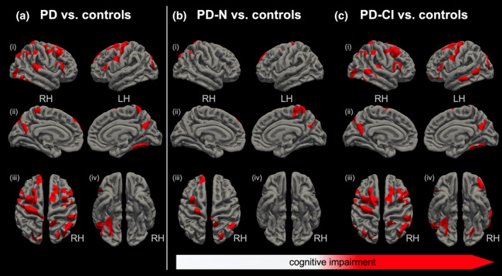
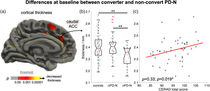
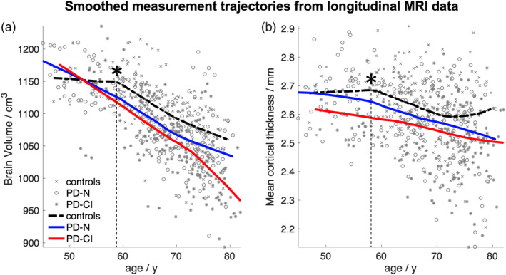
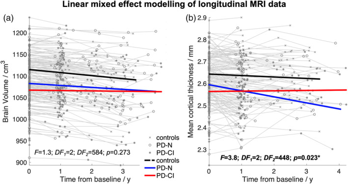
We investigated the brain atrophy distribution pattern and rate of regional atrophy change in Parkinson's disease (PD) in association with the cognitive status to identify the morphological characteristics of conversion to mild cognitive impairment (MCI) and dementia (PDD). T1-weighted longitudinal 3T MRI data (up to four follow-up assessments) from neuropsychologically well-characterized advanced PD patients (n = 172, 8.9 years disease duration) and healthy elderly controls (n = 85) enrolled in the LANDSCAPE study were longitudinally analyzed using a linear mixed effect model and atlas-based volumetry and cortical thickness measures. At baseline, PD patients presented with cerebral atrophy and cortical thinning including striatum, temporoparietal regions, and primary/premotor cortex. The atrophy was already observed in "cognitively normal" PD patients (PD-N) and was considerably more pronounced in cognitively impaired PD patients. Linear mixed effect modeling revealed almost similar rates of atrophy change in PD and controls. The group comparison at baseline between those PD-N whose cognitive performance remained stable (n = 42) and those PD-N patients who converted to MCI/PDD ("converter" cPD-N, n = 26) indicated suggested cortical thinning in the anterior cingulate cortex in cPD-N patients which was correlated with cognitive performance. Our results suggest that cortical brain atrophy has been already expanded in advanced PD patients without overt cognitive deficits while atrophy progression in late disease did not differ from "normal" aging regardless of the cognitive status. It appears that cortical atrophy begins early and progresses already in the initial disease stages emphasizing the need for therapeutic interventions already at disease onset.
我们研究了帕金森病(PD)患者脑萎缩的分布模式和区域萎缩变化率与认知状态的关系,以确定向轻度认知障碍(MCI)和痴呆(PDD)转化的形态学特征。对来自认知功能良好的晚期 PD 患者(n=172,疾病病程 8.9 年)和 LANDSCAPE 研究中招募的健康老年对照组(n=85)的 3T MRI 数据(最多 4 次随访评估)进行了纵向分析,采用线性混合效应模型和基于图谱的体积和皮质厚度测量。在基线时,PD 患者表现出脑萎缩和皮质变薄,包括纹状体、颞顶叶区域和初级/运动前皮质。这种萎缩在“认知正常”的 PD 患者(PD-N)中已经观察到,在认知受损的 PD 患者中更为明显。线性混合效应模型显示,PD 患者和对照组的萎缩变化率几乎相似。在基线时,将认知表现稳定的 PD-N 患者(n=42)和那些转为 MCI/PDD 的 PD-N 患者(“转化”cPD-N,n=26)进行组间比较,结果表明 cPD-N 患者的前扣带皮质存在皮质变薄,与认知表现相关。我们的结果表明,在没有明显认知缺陷的晚期 PD 患者中,皮质脑萎缩已经扩大,而晚期疾病的萎缩进展与“正常”衰老没有区别,无论认知状态如何。皮质萎缩似乎很早就开始了,并已在疾病的初始阶段进展,这强调了在疾病发病时就需要进行治疗干预。