Privolzhsky Research Medical University, Minina Square 10/1, 603005, Nizhny Novgorod, Russia.
A. Tsyb Medical Radiological Research Center, Korolev Street 4, Obninsk, 249036, Kaluga region, Russia.
Sci Rep. 2019 Dec 10;9(1):18670. doi: 10.1038/s41598-019-55215-6.
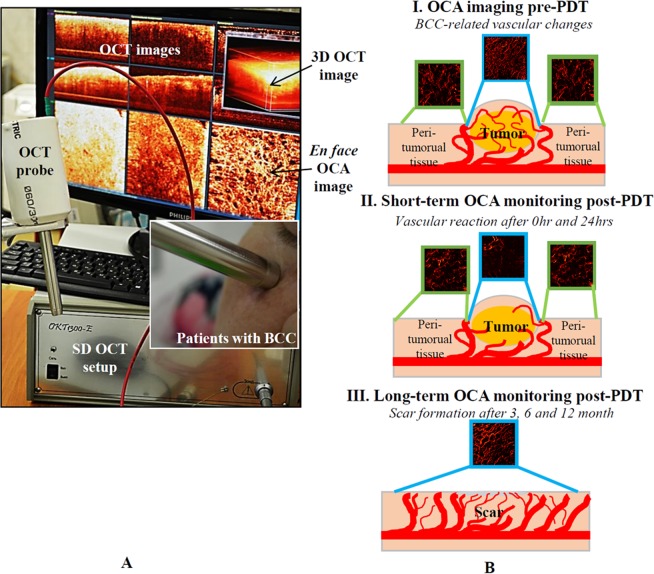
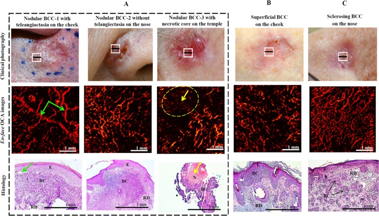
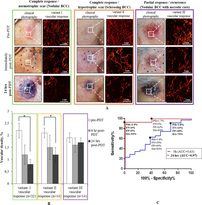
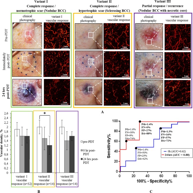
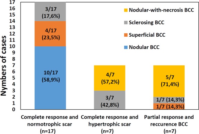
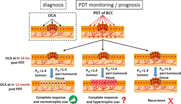
Microvascular networks of human basal cell carcinomas (BCC) and surrounding skin were assessed with optical coherence angiography (OCA) in conjunction with photodynamic therapy (PDT). OCA images were collected and analyzed in 31 lesions pre-treatment, and immediately/24 hours/3-12 months post-treatment. Pre-treatment OCA enabled differentiation between prevalent subtypes of BCC (nodular and superficial) and nodular-with-necrotic-core BCC subtypes with a diagnostic accuracy of 78%; this can facilitate more accurate biopsy reducing sampling error and better therapy regimen selection. Post-treatment OCA images at 24 hours were 98% predictive of eventual outcome. Additional findings highlight the importance of pre-treatment necrotic core, vascular metrics associated with hypertrophic scar formation, and early microvascular changes necessary in both tumorous and peri-tumorous regions to ensure treatment success.
采用光学相干血管造影术(OCA)结合光动力疗法(PDT)评估了人基底细胞癌(BCC)及其周围皮肤的微血管网络。在治疗前,对 31 个病变部位采集和分析了 OCA 图像,并在治疗后即刻/24 小时/3-12 个月进行了分析。治疗前的 OCA 能够区分常见的 BCC 亚型(结节型和浅表型)和伴有坏死核心的结节型 BCC 亚型,其诊断准确性为 78%;这有助于进行更准确的活检,减少采样误差,更好地选择治疗方案。治疗后 24 小时的 OCA 图像对最终结果的预测准确率为 98%。其他发现强调了治疗前坏死核心、与增生性瘢痕形成相关的血管指标以及在肿瘤和肿瘤周围区域中早期微血管变化的重要性,这些变化是确保治疗成功所必需的。