Department Psychology and Neurosciences, Leibniz Research Centre for Working Environment and Human Factors, Dortmund, Germany.
REVAL Research Institute, University of Hasselt, Hasselt, Belgium.
Hum Brain Mapp. 2020 Apr 15;41(6):1644-1666. doi: 10.1002/hbm.24901. Epub 2019 Dec 20.
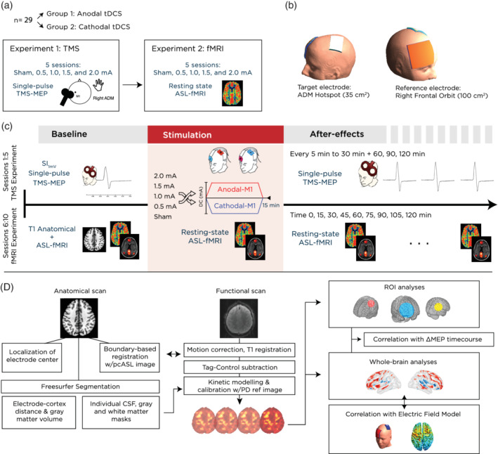
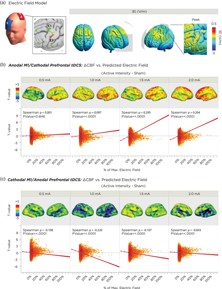
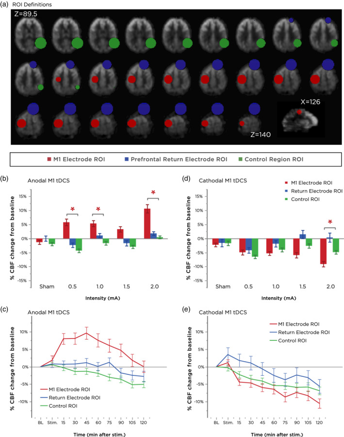
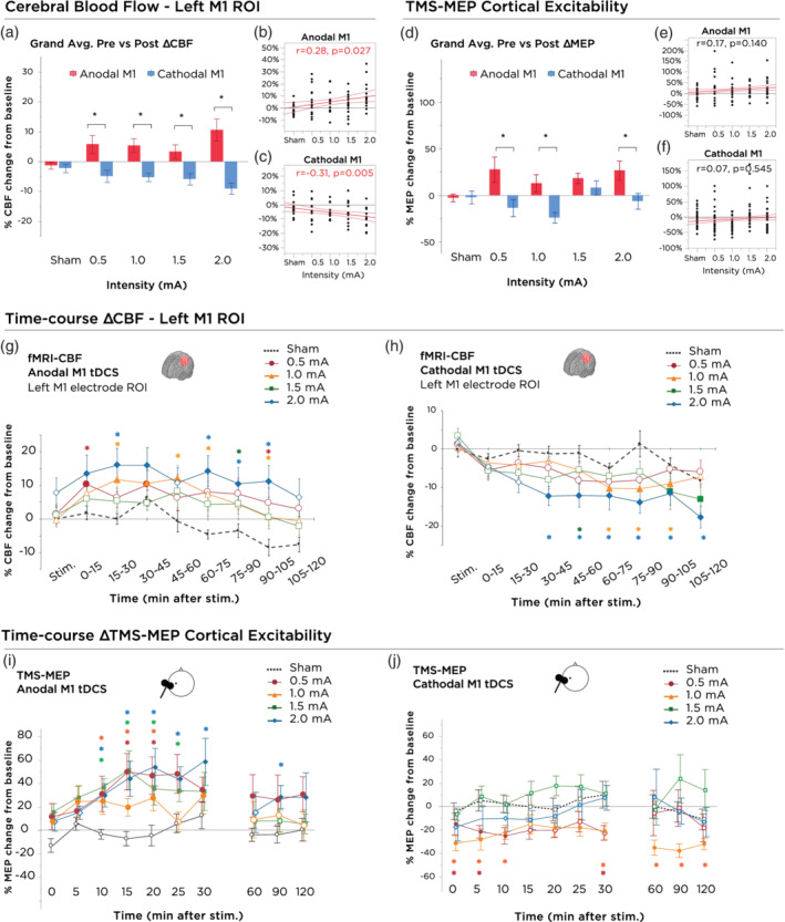
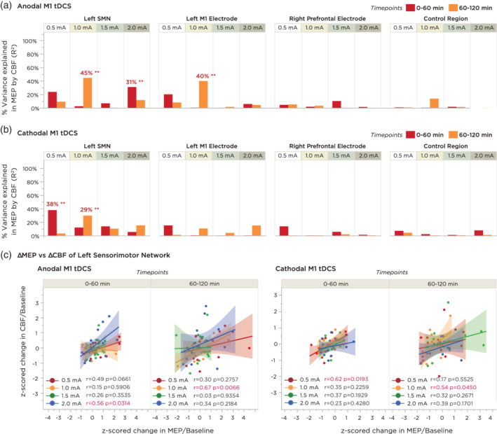
Transcranial direct current stimulation (tDCS) induces polarity- and dose-dependent neuroplastic aftereffects on cortical excitability and cortical activity, as demonstrated by transcranial magnetic stimulation (TMS) and functional imaging (fMRI) studies. However, lacking systematic comparative studies between stimulation-induced changes in cortical excitability obtained from TMS, and cortical neurovascular activity obtained from fMRI, prevent the extrapolation of respective physiological and mechanistic bases. We investigated polarity- and intensity-dependent effects of tDCS on cerebral blood flow (CBF) using resting-state arterial spin labeling (ASL-MRI), and compared the respective changes to TMS-induced cortical excitability (amplitudes of motor evoked potentials, MEP) in separate sessions within the same subjects (n = 29). Fifteen minutes of sham, 0.5, 1.0, 1.5, and 2.0-mA anodal or cathodal tDCS was applied over the left primary motor cortex (M1) in a randomized repeated-measure design. Time-course changes were measured before, during and intermittently up to 120-min after stimulation. ROI analyses indicated linear intensity- and polarity-dependent tDCS after-effects: all anodal-M1 intensities increased CBF under the M1 electrode, with 2.0-mA increasing CBF the greatest (15.3%) compared to sham, while all cathodal-M1 intensities decreased left M1 CBF from baseline, with 2.0-mA decreasing the greatest (-9.3%) from sham after 120-min. The spatial distribution of perfusion changes correlated with the predicted electric field, as simulated with finite element modeling. Moreover, tDCS-induced excitability changes correlated more strongly with perfusion changes in the left sensorimotor region compared to the targeted hand-knob region. Our findings reveal lasting tDCS-induced alterations in cerebral perfusion, which are dose-dependent with tDCS parameters, but only partially account for excitability changes.
经颅直流电刺激(tDCS)通过经颅磁刺激(TMS)和功能成像(fMRI)研究证明,可引起皮质兴奋性和皮质活动的极性和剂量依赖性神经可塑性后效。然而,缺乏 TMS 获得的皮质兴奋性刺激诱导变化与 fMRI 获得的皮质神经血管活性之间的系统比较研究,限制了各自生理和机制基础的推断。我们使用静息状态动脉自旋标记(ASL-MRI)研究了 tDCS 对脑血流(CBF)的极性和强度依赖性影响,并在同一受试者的单独会话中比较了 TMS 诱导的皮质兴奋性(运动诱发电位振幅,MEP)的各自变化(n = 29)。在随机重复测量设计中,在左初级运动皮层(M1)上施加 15 分钟的假刺激、0.5、1.0、1.5 和 2.0 mA 阳极或阴极 tDCS。在刺激之前、期间和间歇性测量到 120 分钟的时间过程变化。ROI 分析表明 tDCS 后效具有线性强度和极性依赖性:所有阳极-M1 强度均增加了 M1 电极下的 CBF,2.0 mA 增加的 CBF 最大(与假刺激相比增加 15.3%),而所有阴极-M1 强度均降低了左 M1 CBF 与基线相比,2.0 mA 在 120 分钟后从假刺激中降低的最大(-9.3%)。灌注变化的空间分布与有限元建模模拟的预测电场相关。此外,与目标手旋钮区域相比,tDCS 诱导的兴奋性变化与左感觉运动区域的灌注变化相关性更强。我们的发现揭示了持久的 tDCS 诱导的脑灌注变化,这些变化与 tDCS 参数呈剂量依赖性,但仅部分解释了兴奋性变化。