Amrollahi Pouya, Rodrigues Meryl, Lyon Christopher J, Goel Ajay, Han Haiyong, Hu Tony Y
Virginia G. Piper Biodesign Center for Personalized Diagnostics, The Biodesign Institute, Arizona State University, Tempe, AZ, United States.
School of Biological and Health Systems Engineering, Arizona State University, Tempe, AZ, United States.
Front Genet. 2019 Dec 17;10:1273. doi: 10.3389/fgene.2019.01273. eCollection 2019.
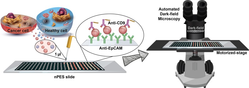
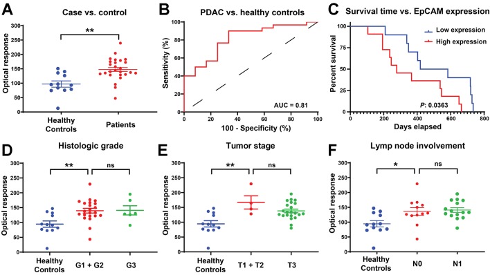
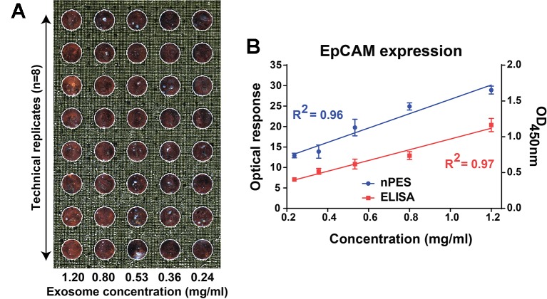
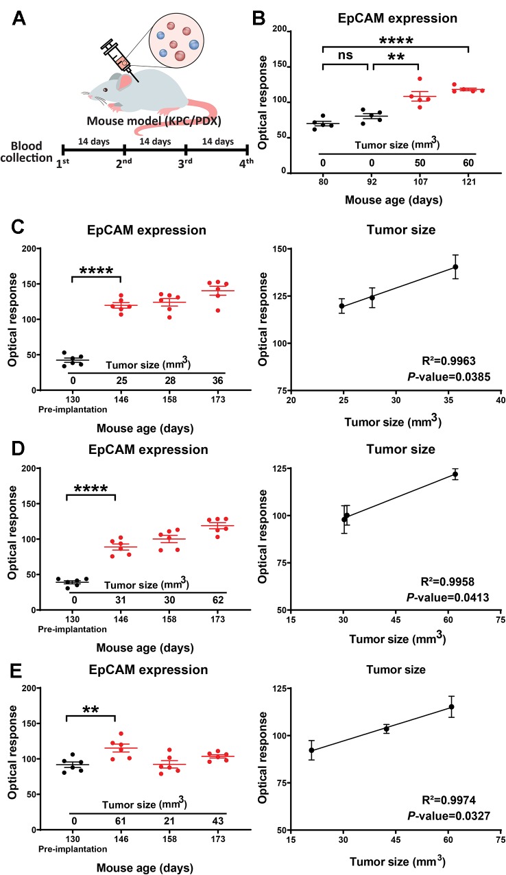
Extracellular vesicles (EVs) are abundant in most biological fluids and considered promising biomarker candidates, but the development of EV biomarker assays is hindered, in part, by their requirement for prior EV purification and the lack of standardized and reproducible EV isolation methods. We now describe a far-field nanoplasmon-enhanced scattering (FF-nPES) assay for the isolation-free characterization of EVs present in small volumes of serum (< 5 µl). In this approach, EVs are captured with a cancer-selective antibody, hybridized with gold nanorods conjugated with an antibody to the EV surface protein CD9, and quantified by their ability to scatter light when analyzed using a fully automated dark-field microscope system. Our results indicate that FF-nPES performs similarly to EV ELISA, when analyzing EV surface expression of epithelial cell adhesion molecule (EpCAM), which has clinical significant as a cancer biomarker. Proof-of-concept FF-nPES data indicate that it can directly analyze EV EpCAM expression from serum samples to distinguish early stage pancreatic ductal adenocarcinoma patients from healthy subjects, detect the development of early stage tumors in a mouse model of spontaneous pancreatic cancer, and monitor tumor growth in patient derived xenograft mouse models of pancreatic cancer. FF-nPES thus appears to exhibit strong potential for the direct analysis of EV membrane biomarkers for disease diagnosis and treatment monitoring.
细胞外囊泡(EVs)在大多数生物体液中含量丰富,被认为是很有前景的生物标志物候选物,但EV生物标志物检测方法的发展受到一定阻碍,部分原因在于其需要预先对EV进行纯化,且缺乏标准化和可重复的EV分离方法。我们现在描述一种远场纳米等离子体增强散射(FF-nPES)检测方法,用于对少量血清(<5微升)中存在的EV进行无需分离的表征。在这种方法中,EVs用癌症选择性抗体捕获,与缀合有针对EV表面蛋白CD9的抗体的金纳米棒杂交,并在使用全自动暗场显微镜系统进行分析时,通过它们散射光的能力进行定量。我们的结果表明,在分析上皮细胞粘附分子(EpCAM)的EV表面表达时,FF-nPES的表现与EV ELISA类似,EpCAM作为癌症生物标志物具有临床意义。概念验证性的FF-nPES数据表明,它可以直接分析血清样本中EV的EpCAM表达,以区分早期胰腺导管腺癌患者和健康受试者,在自发性胰腺癌小鼠模型中检测早期肿瘤的发展,并在胰腺癌患者来源的异种移植小鼠模型中监测肿瘤生长。因此,FF-nPES似乎在直接分析用于疾病诊断和治疗监测的EV膜生物标志物方面具有强大潜力。