Pilania Khushboo, Jankharia Bhavin, Monoot Pradeep
Department of Radiodiagnosis, Jankharia Imaging Centre, 383 S V P Road, Bhaveshwar Vihar, Mumbai, Maharashtra, India.
Department of Orthopaedics, Breach Candy Hospital Trust, 60 A Bhulabhai Desai Road, Girgaon, Mumbai, Maharashtra, India.
Indian J Radiol Imaging. 2019 Oct-Dec;29(4):364-371. doi: 10.4103/ijri.IJRI_288_19. Epub 2019 Dec 31.
Till date, weight-bearing radiographs have been the cornerstone for planning surgeries on flatfoot. The technique, however, has limitations due to the superimposition of the bones and the lack of reproducibility. Weight-bearing CT with its unique design overcomes these limitations and enables cross-sectional imaging of the foot to be done in the natural weight-bearing position. In this paper, we report our initial experience in weight-bearing cross-sectional imaging of the foot for assessment of flatfoot deformity.
Around 19 known cases of flatfoot were scanned on the weight-bearing CT. Each foot was then assessed for the various angles and also for the presence/absence of extra-articular talocalcaneal impingement and subfibular impingement. Other associated abnormalities like secondary osteoarthritic changes, were also noted.
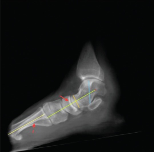
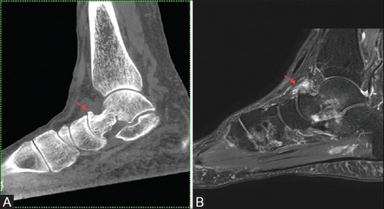
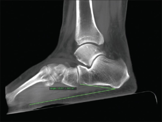
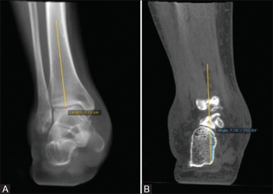
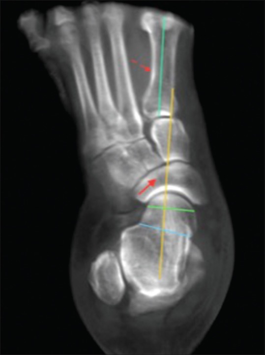
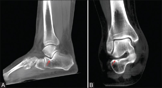
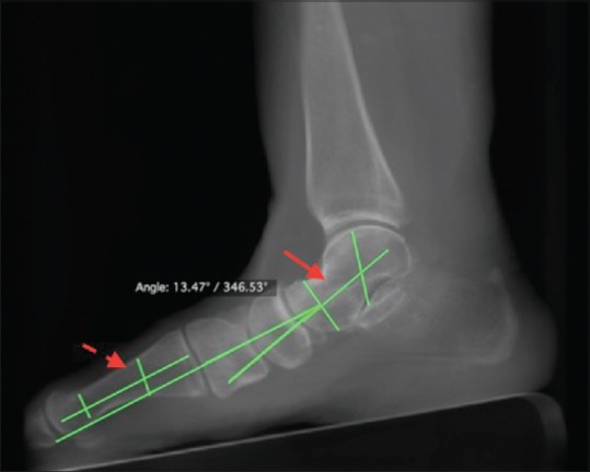
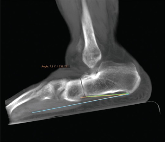
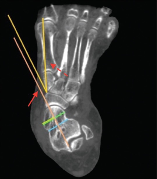
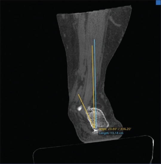
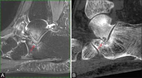
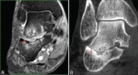
The Meary, as well as the calcaneal angles, were abnormal, in all but one separate foot. Forefoot abduction was seen in 7 of the 19 feet. The hind foot valgus angle was greater than 10° in all patients. Extra-articular talocalcaneal impingement was seen in 13 of 19 feet. Secondary osteoarthritic changes were seen in 14 feet.
Weight-bearing CT scan is a very useful technique for evaluation of flatfoot and associated complications. It overcomes the limitations of the radiographs by providing multiplanar three-dimensional assessment of the foot in the natural weight-bearing position and at the same time being easily reproducible and consistent for the measurements around the foot. The definite advantage over the conventional cross-sectional scanners is the weight-bearing capability.
迄今为止,负重X线片一直是扁平足手术规划的基石。然而,由于骨骼的重叠以及缺乏可重复性,该技术存在局限性。负重CT凭借其独特的设计克服了这些局限性,并能够在自然负重位置对足部进行横断面成像。在本文中,我们报告了我们在负重横断面成像评估扁平足畸形方面的初步经验。
对约19例已知的扁平足病例进行负重CT扫描。然后对每只脚进行各种角度的评估,以及评估是否存在关节外距跟撞击和腓骨下撞击。还记录了其他相关异常,如继发性骨关节炎改变。
除一只单独的脚外,所有脚的Meary角以及跟骨角均异常。19只脚中有7只出现前足外展。所有患者的后足外翻角均大于10°。19只脚中有13只出现关节外距跟撞击。14只脚出现继发性骨关节炎改变。
负重CT扫描是评估扁平足及相关并发症的一项非常有用的技术。它通过在自然负重位置对足部进行多平面三维评估克服了X线片的局限性,同时对于足部周围的测量易于重复且结果一致。相对于传统横断面扫描仪的明确优势在于其负重能力。