• 文献检索
  • 文档翻译
  • 深度研究
  • 学术资讯
  • Suppr Zotero 插件Zotero 插件
  • 邀请有礼
  • 套餐&价格
  • 历史记录
应用&插件
Suppr Zotero 插件Zotero 插件浏览器插件Mac 客户端Windows 客户端微信小程序
定价
高级版会员购买积分包购买API积分包
服务
文献检索文档翻译深度研究API 文档MCP 服务
关于我们
关于 Suppr公司介绍联系我们用户协议隐私条款
关注我们

Suppr 超能文献

核心技术专利:CN118964589B侵权必究
粤ICP备2023148730 号-1Suppr @ 2026

文献检索

告别复杂PubMed语法,用中文像聊天一样搜索,搜遍4000万医学文献。AI智能推荐,让科研检索更轻松。

立即免费搜索

文件翻译

保留排版,准确专业,支持PDF/Word/PPT等文件格式,支持 12+语言互译。

免费翻译文档

深度研究

AI帮你快速写综述,25分钟生成高质量综述,智能提取关键信息,辅助科研写作。

立即免费体验

对人类MICAL3中含黄素单加氧酶和钙调蛋白同源结构域的结构与动力学见解。

Structural and kinetic insights into flavin-containing monooxygenase and calponin-homology domains in human MICAL3.

作者信息

Kim Junsoo, Lee Haemin, Roh Yeon Jin, Kim Han-Ul, Shin Donghyuk, Kim Sorah, Son Jonghyeon, Lee Aro, Kim Minseo, Park Junga, Hwang Seong Yun, Kim Kyunghwan, Lee Yong Kwon, Jung Hyun Suk, Hwang Kwang Yeon, Lee Byung Cheon

机构信息

College of Life Sciences and Biotechnology, Korea University, 145 Anam-ro, Seongbuk-gu, Seoul 02841, Republic of Korea.

Biochemistry Laboratory, Department of Biosystems and Biotechnology, Kangwon National University, 1 Kangwondaekak-gil, Chuncheon-si, Gangwon-do 24341, Republic of Korea.

出版信息

IUCrJ. 2020 Jan 1;7(Pt 1):90-99. doi: 10.1107/S2052252519015409.

DOI:10.1107/S2052252519015409
PMID:31949908
原文链接:https://pmc.ncbi.nlm.nih.gov/articles/PMC6949599/
Abstract

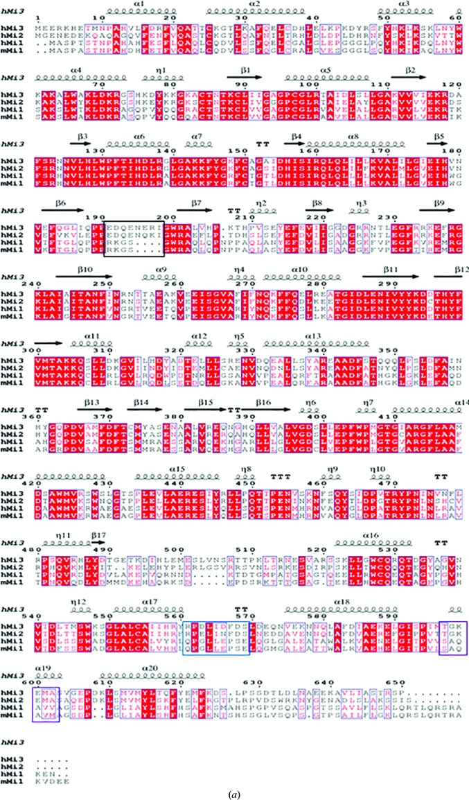
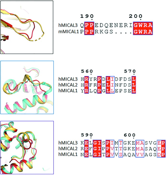
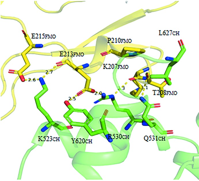
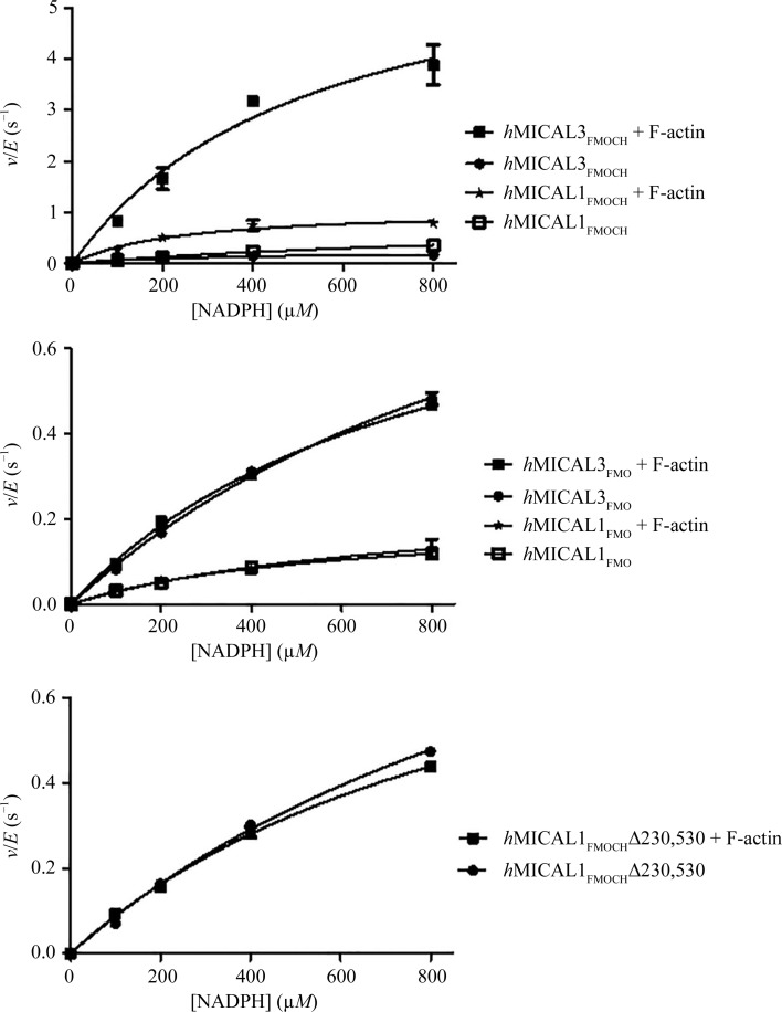
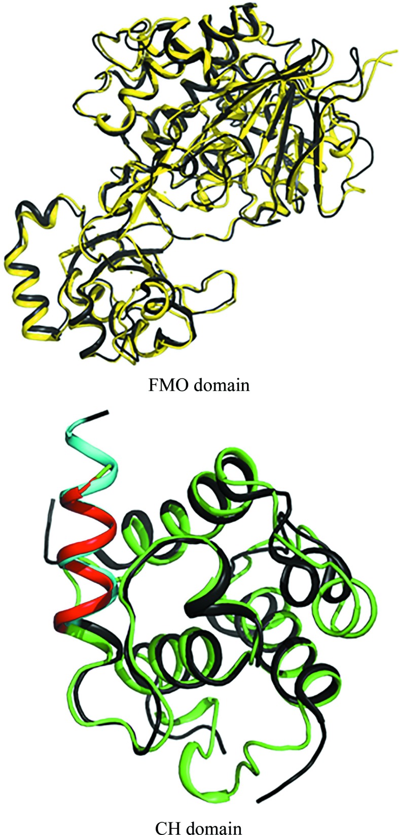
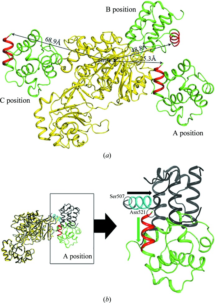
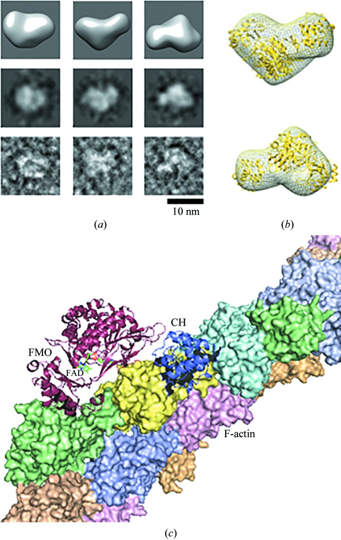
MICAL is an oxidoreductase that participates in cytoskeleton reorganization via actin disassembly in the presence of NADPH. Although three MICALs (MICAL1, MICAL2 and MICAL3) have been identified in mammals, only the structure of mouse MICAL1 has been reported. Here, the first crystal structure of human MICAL3, which contains the flavin-containing monooxygenase (FMO) and calponin-homology (CH) domains, is reported. MICAL3 has an FAD/NADP-binding Rossmann-fold domain for mono-oxygenase activity like MICAL1. The FMO and CH domains of both MICAL3 and MICAL1 are highly similar in structure, but superimposition of the two structures shows a different relative position of the CH domain in the asymmetric unit. Based on kinetic analyses, the catalytic efficiency of MICAL3 dramatically increased on adding F-actin only when the CH domain was available. However, this did not occur when two residues, Glu213 and Arg530, were mutated in the FMO and CH domains, respectively. Overall, MICAL3 is structurally highly similar to MICAL1, which suggests that they may adopt the same catalytic mechanism, but the difference in the relative position of the CH domain produces a difference in F-actin substrate specificity.

摘要

MICAL是一种氧化还原酶,在NADPH存在的情况下,通过肌动蛋白解聚参与细胞骨架重组。尽管在哺乳动物中已鉴定出三种MICAL(MICAL1、MICAL2和MICAL3),但仅报道了小鼠MICAL1的结构。在此,报道了人MICAL3的首个晶体结构,其包含含黄素单加氧酶(FMO)和卡波宁同源(CH)结构域。MICAL3具有一个用于单加氧酶活性的FAD/NADP结合罗斯曼折叠结构域,与MICAL1类似。MICAL3和MICAL1的FMO和CH结构域在结构上高度相似,但两种结构的叠加显示CH结构域在不对称单元中的相对位置不同。基于动力学分析,仅当CH结构域存在时,添加F-肌动蛋白会使MICAL3的催化效率显著提高。然而,当FMO和CH结构域中的两个残基分别为Glu213和Arg530突变时,情况并非如此。总体而言,MICAL3在结构上与MICAL1高度相似,这表明它们可能采用相同的催化机制,但CH结构域相对位置的差异导致了F-肌动蛋白底物特异性的差异。

https://cdn.ncbi.nlm.nih.gov/pmc/blobs/5e0f/6949599/ffaebc2a79af/m-07-00090-fig7.jpg
https://cdn.ncbi.nlm.nih.gov/pmc/blobs/5e0f/6949599/d754dc5d6dab/m-07-00090-fig1a.jpg
https://cdn.ncbi.nlm.nih.gov/pmc/blobs/5e0f/6949599/609c8f0a6f10/m-07-00090-fig2.jpg
https://cdn.ncbi.nlm.nih.gov/pmc/blobs/5e0f/6949599/04c7eed5456a/m-07-00090-fig3.jpg
https://cdn.ncbi.nlm.nih.gov/pmc/blobs/5e0f/6949599/a11d47f4be2c/m-07-00090-fig4.jpg
https://cdn.ncbi.nlm.nih.gov/pmc/blobs/5e0f/6949599/9497a3f20094/m-07-00090-fig5.jpg
https://cdn.ncbi.nlm.nih.gov/pmc/blobs/5e0f/6949599/896ccb890c65/m-07-00090-fig6.jpg
https://cdn.ncbi.nlm.nih.gov/pmc/blobs/5e0f/6949599/ffaebc2a79af/m-07-00090-fig7.jpg

相似文献

1
Structural and kinetic insights into flavin-containing monooxygenase and calponin-homology domains in human MICAL3.对人类MICAL3中含黄素单加氧酶和钙调蛋白同源结构域的结构与动力学见解。
IUCrJ. 2020 Jan 1;7(Pt 1):90-99. doi: 10.1107/S2052252519015409.
2
Properties and catalytic activities of MICAL1, the flavoenzyme involved in cytoskeleton dynamics, and modulation by its CH, LIM and C-terminal domains.参与细胞骨架动力学的黄素酶MICAL1的特性与催化活性及其CH、LIM和C末端结构域的调节作用
Arch Biochem Biophys. 2016 Mar 1;593:24-37. doi: 10.1016/j.abb.2016.01.016. Epub 2016 Feb 1.
3
Enhanced Production of the Mical Redox Domain for Enzymology and F-actin Disassembly Assays.增强微管相关蛋白轻链(Mical)氧化还原结构域用于酶学和 F-actin 解聚分析。
Int J Mol Sci. 2021 Feb 17;22(4):1991. doi: 10.3390/ijms22041991.
4
Modulation of MICAL Monooxygenase Activity by its Calponin Homology Domain: Structural and Mechanistic Insights.钙调蛋白同源结构域对MICAL单加氧酶活性的调节:结构与机制解析
Sci Rep. 2016 Mar 3;6:22176. doi: 10.1038/srep22176.
5
Autoinhibition and relief mechanisms for MICAL monooxygenases in F-actin disassembly.微管相关蛋白轻链 3 单加氧酶在 F-actin 解聚中的自动抑制和缓解机制。
Nat Commun. 2024 Aug 9;15(1):6824. doi: 10.1038/s41467-024-50940-7.
6
Differential regulation of actin microfilaments by human MICAL proteins.人 MICAL 蛋白对肌动蛋白微丝的差异调节。
J Cell Sci. 2012 Feb 1;125(Pt 3):614-24. doi: 10.1242/jcs.089367. Epub 2012 Feb 13.
7
MICAL-mediated oxidation of actin and its effects on cytoskeletal and cellular dynamics.MICAL介导的肌动蛋白氧化及其对细胞骨架和细胞动力学的影响。
Front Cell Dev Biol. 2023 Feb 17;11:1124202. doi: 10.3389/fcell.2023.1124202. eCollection 2023.
8
Human MICAL1: Activation by the small GTPase Rab8 and small-angle X-ray scattering studies on the oligomerization state of MICAL1 and its complex with Rab8.人 MICAL1:由小分子 GTPase Rab8 激活及其与 Rab8 复合物的寡聚状态的小角 X 射线散射研究。
Protein Sci. 2019 Jan;28(1):150-166. doi: 10.1002/pro.3512. Epub 2018 Oct 31.
9
Actin stimulates reduction of the MICAL-2 monooxygenase domain.肌动蛋白刺激 MICAL-2 单加氧酶结构域的还原。
Biochemistry. 2013 Sep 3;52(35):6076-84. doi: 10.1021/bi4008462. Epub 2013 Aug 21.
10
MICAL1 activation by PAK1 mediates actin filament disassembly.PAK1 介导的 MICAL1 激活介导肌动蛋白丝解体。
Cell Rep. 2022 Oct 4;41(1):111442. doi: 10.1016/j.celrep.2022.111442.

引用本文的文献

1
Structural basis of MICAL autoinhibition.微管相关蛋白轻链(MICAL)自动抑制的结构基础。
Nat Commun. 2024 Nov 12;15(1):9810. doi: 10.1038/s41467-024-54131-2.
2
CHK1 controls zygote pronuclear envelope breakdown by regulating F-actin through interacting with MICAL3.chk1 通过与 mical3 相互作用来调节 f-actin 控制合子原核膜破裂。
EMBO Rep. 2024 Nov;25(11):4876-4897. doi: 10.1038/s44319-024-00267-7. Epub 2024 Oct 2.
3
Autoinhibition and relief mechanisms for MICAL monooxygenases in F-actin disassembly.微管相关蛋白轻链 3 单加氧酶在 F-actin 解聚中的自动抑制和缓解机制。

本文引用的文献

1
The MICALs are a Family of F-actin Dismantling Oxidoreductases Conserved from Drosophila to Humans.微管相关蛋白轻链(MICALs)是一类从果蝇到人类都保守的 F-actin 解聚氧化还原酶家族。
Sci Rep. 2018 Jan 17;8(1):937. doi: 10.1038/s41598-017-17943-5.
2
PyMod 2.0: improvements in protein sequence-structure analysis and homology modeling within PyMOL.PyMod 2.0:PyMOL 中蛋白质序列-结构分析和同源建模的改进。
Bioinformatics. 2017 Feb 1;33(3):444-446. doi: 10.1093/bioinformatics/btw638.
3
Processing of X-ray diffraction data collected in oscillation mode.
Nat Commun. 2024 Aug 9;15(1):6824. doi: 10.1038/s41467-024-50940-7.
4
MICAL-mediated oxidation of actin and its effects on cytoskeletal and cellular dynamics.MICAL介导的肌动蛋白氧化及其对细胞骨架和细胞动力学的影响。
Front Cell Dev Biol. 2023 Feb 17;11:1124202. doi: 10.3389/fcell.2023.1124202. eCollection 2023.
5
Monooxygenase in Autosomal Dominant Lateral Temporal Epilepsy: Role in Cytoskeletal Regulation and Relation to Cancer.常染色体显性颞叶癫痫的单加氧酶:细胞骨架调节中的作用及其与癌症的关系。
Genes (Basel). 2022 Apr 19;13(5):715. doi: 10.3390/genes13050715.
6
Structural Characteristics, Binding Partners and Related Diseases of the Calponin Homology (CH) Domain.钙调蛋白同源(CH)结构域的结构特征、结合伴侣及相关疾病
Front Cell Dev Biol. 2020 May 14;8:342. doi: 10.3389/fcell.2020.00342. eCollection 2020.
振荡模式下收集的X射线衍射数据的处理。
Methods Enzymol. 1997;276:307-26. doi: 10.1016/S0076-6879(97)76066-X.
4
Single-Particle Refinement and Variability Analysis in EMAN2.1.EMAN2.1中的单颗粒精修与变异性分析
Methods Enzymol. 2016;579:159-89. doi: 10.1016/bs.mie.2016.05.001. Epub 2016 Jul 1.
5
Modulation of MICAL Monooxygenase Activity by its Calponin Homology Domain: Structural and Mechanistic Insights.钙调蛋白同源结构域对MICAL单加氧酶活性的调节:结构与机制解析
Sci Rep. 2016 Mar 3;6:22176. doi: 10.1038/srep22176.
6
Regulation of protein function by reversible methionine oxidation and the role of selenoprotein MsrB1.可逆蛋氨酸氧化对蛋白质功能的调控及硒蛋白MsrB1的作用
Antioxid Redox Signal. 2015 Oct 1;23(10):814-22. doi: 10.1089/ars.2015.6385. Epub 2015 Jul 16.
7
Redox modification of nuclear actin by MICAL-2 regulates SRF signaling.MICAL-2 通过氧化还原修饰核肌动蛋白调节 SRF 信号转导。
Cell. 2014 Jan 30;156(3):563-76. doi: 10.1016/j.cell.2013.12.035. Epub 2014 Jan 16.
8
The physiological role of reversible methionine oxidation.可逆性甲硫氨酸氧化的生理作用。
Biochim Biophys Acta. 2014 Aug;1844(8):1367-82. doi: 10.1016/j.bbapap.2014.01.001. Epub 2014 Jan 10.
9
MsrB1 and MICALs regulate actin assembly and macrophage function via reversible stereoselective methionine oxidation.MsrB1 和 MICALs 通过可逆立体选择性蛋氨酸氧化调节肌动蛋白组装和巨噬细胞功能。
Mol Cell. 2013 Aug 8;51(3):397-404. doi: 10.1016/j.molcel.2013.06.019. Epub 2013 Aug 1.
10
MICAL-family proteins: Complex regulators of the actin cytoskeleton.MICAL家族蛋白:肌动蛋白细胞骨架的复杂调节因子。
Antioxid Redox Signal. 2014 May 1;20(13):2059-73. doi: 10.1089/ars.2013.5487. Epub 2013 Aug 17.