• 文献检索
  • 文档翻译
  • 深度研究
  • 学术资讯
  • Suppr Zotero 插件Zotero 插件
  • 邀请有礼
  • 套餐&价格
  • 历史记录
应用&插件
Suppr Zotero 插件Zotero 插件浏览器插件Mac 客户端Windows 客户端微信小程序
定价
高级版会员购买积分包购买API积分包
服务
文献检索文档翻译深度研究API 文档MCP 服务
关于我们
关于 Suppr公司介绍联系我们用户协议隐私条款
关注我们

Suppr 超能文献

核心技术专利:CN118964589B侵权必究
粤ICP备2023148730 号-1Suppr @ 2026

文献检索

告别复杂PubMed语法,用中文像聊天一样搜索,搜遍4000万医学文献。AI智能推荐,让科研检索更轻松。

立即免费搜索

文件翻译

保留排版,准确专业,支持PDF/Word/PPT等文件格式,支持 12+语言互译。

免费翻译文档

深度研究

AI帮你快速写综述,25分钟生成高质量综述,智能提取关键信息,辅助科研写作。

立即免费体验

同步放化疗后鼻咽癌患者颞叶微结构异常的高分辨率扩散加权成像和扩散峰度成像定量评估

Temporal lobe microstructural abnormalities in patients with nasopharyngeal carcinoma quantitatively evaluated by high-resolution DWI and DKI after concurrent chemoradiotherapy.

作者信息

Wu Gang, Li Rui-Rui, Balasubramanian Priya S, Li Meng-Meng, Yang Kai, Huang Wei-Yuan, Chen Feng

机构信息

Department of Radiation Oncology, Hainan General Hospital (Hainan Affiliated Hospital of Hainan Medical University), Haikou, China.

Department of Radiology, Hainan Hospital of Hainan Medical College (Hainan General Hospital), Haikou, China.

出版信息

Clin Transl Radiat Oncol. 2019 Dec 25;21:36-43. doi: 10.1016/j.ctro.2019.12.003. eCollection 2020 Mar.

DOI:10.1016/j.ctro.2019.12.003
PMID:31970294
原文链接:https://pmc.ncbi.nlm.nih.gov/articles/PMC6965203/
Abstract

PURPOSE

To investigate temporal lobe microstructural abnormalities and neurocognitive function impairment after concurrent chemoradiotherapy (CCRT) in patients with nasopharyngeal carcinoma (NPC).

METHODS

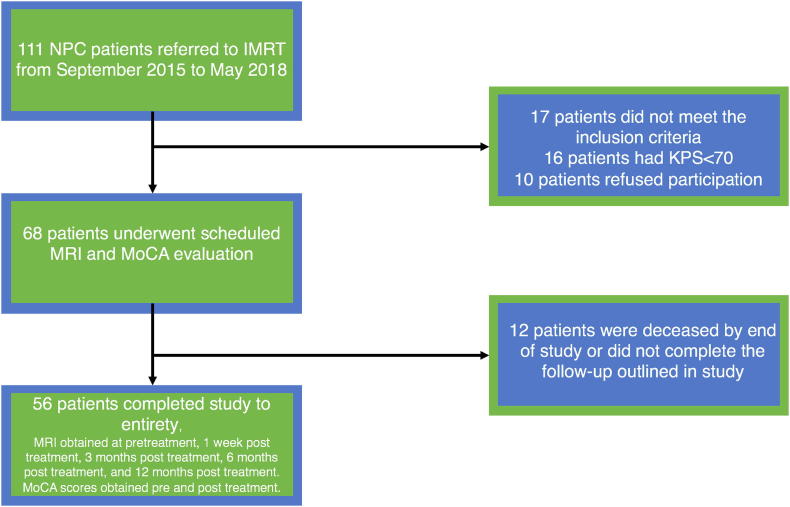
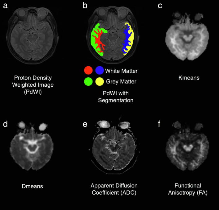
NPC patients who underwent CCRT were enrolled. High-resolution diffusion-weighted imaging (DWI) magnetic resonance imaging (MRI) and diffusion-kurtosis imaging (DKI) MRI, were performed 5 times per patient (once pre-CCRT, 1 week post-CCRT, 3 months post-CCRT, 6 months post-CCRT, and 12 months post-CCRT). Neurocognitive function was evaluated by Montreal Neurocognitive Assessment (MoCA) twice per patient, once pre-CCRT, and once 12-months after CCRT.

RESULTS

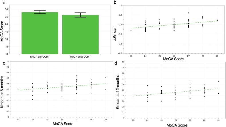
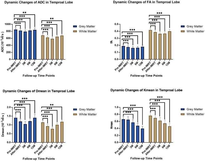
Of 111 patients, 56 completed the entire protocol. The MRI derived apparent diffusion coefficient (ADC), mean of diffusion coefficient (Dmean) and fractional anisotropy (FA) values were significantly decreased ( < 0.05) over the 0-3 month period following CCRT and significantly increased ( < 0.05) over the 3-12 month period following CCRT. The mean of kurtosis coefficient (Kmean) continued to decline over a year post-CCRT. All parameters reveal more pronounced changes in white matter (WM) than in grey matter (GM). MoCA also declined after CCRT ( < 0.001). MoCA showed significant positive correlation with Kmean-WM-6 m, Kmean-WM-12 m and ΔKmean-WM.

CONCLUSIONS

High-resolution DWI and DKI should be considered as a promising method for the investigation of temporal lobe microstructural change in NPC patients after CCRT.

摘要

目的

探讨鼻咽癌(NPC)患者同步放化疗(CCRT)后颞叶微观结构异常及神经认知功能损害情况。

方法

纳入接受CCRT的NPC患者。每位患者进行5次高分辨率扩散加权成像(DWI)磁共振成像(MRI)和扩散峰度成像(DKI)MRI检查(CCRT前1次、CCRT后1周、CCRT后3个月、CCRT后6个月、CCRT后12个月各1次)。每位患者通过蒙特利尔神经认知评估(MoCA)对神经认知功能进行两次评估,一次在CCRT前,一次在CCRT后12个月。

结果

111例患者中,56例完成了整个研究方案。MRI得出的表观扩散系数(ADC)、扩散系数平均值(Dmean)和分数各向异性(FA)值在CCRT后的0 - 3个月期间显著降低(<0.05),在CCRT后的3 - 12个月期间显著升高(<0.05)。峰度系数平均值(Kmean)在CCRT后一年持续下降。所有参数显示白质(WM)的变化比灰质(GM)更明显。CCRT后MoCA评分也下降了(<0.001)。MoCA与Kmean - WM - 6m、Kmean - WM - 12m和ΔKmean - WM呈显著正相关。

结论

高分辨率DWI和DKI应被视为研究NPC患者CCRT后颞叶微观结构变化的一种有前景的方法。

https://cdn.ncbi.nlm.nih.gov/pmc/blobs/33cd/6965203/4c94458d594d/gr4.jpg
https://cdn.ncbi.nlm.nih.gov/pmc/blobs/33cd/6965203/6bae053eb56a/gr1.jpg
https://cdn.ncbi.nlm.nih.gov/pmc/blobs/33cd/6965203/3c89e416cc5b/gr2.jpg
https://cdn.ncbi.nlm.nih.gov/pmc/blobs/33cd/6965203/faf6b63289b3/gr3.jpg
https://cdn.ncbi.nlm.nih.gov/pmc/blobs/33cd/6965203/4c94458d594d/gr4.jpg
https://cdn.ncbi.nlm.nih.gov/pmc/blobs/33cd/6965203/6bae053eb56a/gr1.jpg
https://cdn.ncbi.nlm.nih.gov/pmc/blobs/33cd/6965203/3c89e416cc5b/gr2.jpg
https://cdn.ncbi.nlm.nih.gov/pmc/blobs/33cd/6965203/faf6b63289b3/gr3.jpg
https://cdn.ncbi.nlm.nih.gov/pmc/blobs/33cd/6965203/4c94458d594d/gr4.jpg

相似文献

1
Temporal lobe microstructural abnormalities in patients with nasopharyngeal carcinoma quantitatively evaluated by high-resolution DWI and DKI after concurrent chemoradiotherapy.同步放化疗后鼻咽癌患者颞叶微结构异常的高分辨率扩散加权成像和扩散峰度成像定量评估
Clin Transl Radiat Oncol. 2019 Dec 25;21:36-43. doi: 10.1016/j.ctro.2019.12.003. eCollection 2020 Mar.
2
Early Stage Markers of Late Delayed Neurocognitive Decline Using Diffusion Kurtosis Imaging of Temporal Lobe in Nasopharyngeal Carcinoma Patients.利用鼻咽癌患者颞叶扩散峰度成像检测晚期延迟神经认知功能下降的早期标志物
J Cancer. 2020 Aug 25;11(20):6168-6177. doi: 10.7150/jca.48759. eCollection 2020.
3
Diffusion kurtosis imaging study on temporal lobe after nasopharyngeal carcinoma radiotherapy.鼻咽癌放疗后颞叶的扩散峰度成像研究
Brain Res. 2016 Oct 1;1648(Pt A):387-393. doi: 10.1016/j.brainres.2016.07.041. Epub 2016 Aug 8.
4
Diffusion Kurtosis as an in vivo Imaging Marker of Early Radiation-Induced Changes in Radiation-Induced Temporal Lobe Necrosis in Nasopharyngeal Carcinoma Patients.扩散峰度成像作为鼻咽癌患者放射性颞叶坏死早期辐射诱导变化的一种活体成像标志物
Clin Neuroradiol. 2018 Sep;28(3):413-420. doi: 10.1007/s00062-017-0585-9. Epub 2017 Apr 26.
5
In Vivo Imaging Markers for Prediction of Radiotherapy Response in Patients with Nasopharyngeal Carcinoma: RESOLVE DWI versus DKI.体内影像学标志物预测鼻咽癌放疗反应:RESOLVE DWI 与 DKI 比较。
Sci Rep. 2018 Oct 26;8(1):15861. doi: 10.1038/s41598-018-34072-9.
6
Correlations Between the Microstructural Changes of the Medial Temporal Cortex and Mild Cognitive Impairment in Patients With Cerebral Small Vascular Disease (cSVD): A Diffusion Kurtosis Imaging Study.脑小血管病(cSVD)患者内侧颞叶皮质微观结构变化与轻度认知障碍的相关性:扩散峰度成像研究
Front Neurol. 2020 Jan 15;10:1378. doi: 10.3389/fneur.2019.01378. eCollection 2019.
7
Diffusion-kurtosis imaging predicts early radiotherapy response in nasopharyngeal carcinoma patients.扩散峰度成像可预测鼻咽癌患者的早期放疗反应。
Oncotarget. 2017 Aug 2;8(39):66128-66136. doi: 10.18632/oncotarget.19820. eCollection 2017 Sep 12.
8
Characterization of Alzheimer's Disease Using Ultra-high b-values Apparent Diffusion Coefficient and Diffusion Kurtosis Imaging.使用超高b值表观扩散系数和扩散峰度成像对阿尔茨海默病进行特征描述。
Aging Dis. 2019 Oct 1;10(5):1026-1036. doi: 10.14336/AD.2018.1129. eCollection 2019 Oct.
9
The diffusion-weighted magnetic resonance imaging (DWI) predicts the early response of esophageal squamous cell carcinoma to concurrent chemoradiotherapy.扩散加权磁共振成像(DWI)可预测食管鳞状细胞癌对同步放化疗的早期反应。
Radiother Oncol. 2016 Nov;121(2):246-251. doi: 10.1016/j.radonc.2016.10.021. Epub 2016 Nov 9.
10
Apparent diffusion coefficient histogram shape analysis for monitoring early response in patients with advanced cervical cancers undergoing concurrent chemo-radiotherapy.表观扩散系数直方图形状分析用于监测晚期宫颈癌同步放化疗患者的早期反应
Radiat Oncol. 2016 Oct 22;11(1):141. doi: 10.1186/s13014-016-0715-6.

引用本文的文献

1
Hippocampus sparing volumetric modulated arc therapy in patients with loco-regionally advanced oropharyngeal cancer.局部区域晚期口咽癌患者的海马保护容积调强弧形放疗
Phys Imaging Radiat Oncol. 2022 Sep 24;24:71-75. doi: 10.1016/j.phro.2022.09.008. eCollection 2022 Oct.
2
A systematic review on the use of quantitative imaging to detect cancer therapy adverse effects in normal-appearing brain tissue.系统评价使用定量成像技术检测正常外观脑组织中癌症治疗不良反应的研究。
MAGMA. 2022 Feb;35(1):163-186. doi: 10.1007/s10334-021-00985-2. Epub 2021 Dec 17.
3
Early Stage Markers of Late Delayed Neurocognitive Decline Using Diffusion Kurtosis Imaging of Temporal Lobe in Nasopharyngeal Carcinoma Patients.

本文引用的文献

1
Bevacizumab is more effective in nasopharyngeal carcinoma patients with lower maximum radiation dose to the temporal lobe.贝伐单抗对颞叶最大放射剂量较低的鼻咽癌患者更有效。
Chin Clin Oncol. 2019 Oct;8(S1):S20. doi: 10.21037/cco.2019.02.06. Epub 2019 Apr 17.
2
A cross sectional study in cognitive and neurobehavioral impairment in long-term nasopharyngeal cancer survivors treated with intensity-modulated radiotherapy.调强放疗后长期生存的鼻咽癌患者认知和神经行为损伤的横断面研究。
Radiother Oncol. 2019 Feb;131:179-185. doi: 10.1016/j.radonc.2018.09.012. Epub 2018 Sep 29.
3
10-Year Results of Therapeutic Ratio by Intensity-Modulated Radiotherapy Versus Two-Dimensional Radiotherapy in Patients with Nasopharyngeal Carcinoma.
利用鼻咽癌患者颞叶扩散峰度成像检测晚期延迟神经认知功能下降的早期标志物
J Cancer. 2020 Aug 25;11(20):6168-6177. doi: 10.7150/jca.48759. eCollection 2020.
4
Image guidance in radiation therapy for better cure of cancer.放射治疗中的图像引导以更好地治愈癌症。
Mol Oncol. 2020 Jul;14(7):1470-1491. doi: 10.1002/1878-0261.12751. Epub 2020 Jun 29.
调强放疗与二维放疗治疗鼻咽癌患者的 10 年疗效比。
Oncologist. 2019 Jan;24(1):e38-e45. doi: 10.1634/theoncologist.2017-0577. Epub 2018 Aug 6.
4
Longitudinal brain structural alterations in patients with nasopharyngeal carcinoma early after radiotherapy.鼻咽癌放疗后早期患者的脑结构纵向改变。
Neuroimage Clin. 2018 Apr 23;19:252-259. doi: 10.1016/j.nicl.2018.04.019. eCollection 2018.
5
Clinical applications of diffusion weighted imaging in neuroradiology.扩散加权成像在神经放射学中的临床应用。
Insights Imaging. 2018 Aug;9(4):535-547. doi: 10.1007/s13244-018-0624-3. Epub 2018 May 30.
6
Pretreatment neurocognitive function and self-reported symptoms in patients with newly diagnosed head and neck cancer compared with noncancer cohort.新发头颈部癌症患者与非癌症队列患者的预处理神经认知功能和自我报告症状比较。
Head Neck. 2018 Sep;40(9):2029-2042. doi: 10.1002/hed.25198. Epub 2018 Apr 17.
7
Association of Neurocognitive Deficits With Radiotherapy or Chemoradiotherapy for Patients With Head and Neck Cancer.头颈部癌患者神经认知缺陷与放射治疗或放化疗的关联
JAMA Otolaryngol Head Neck Surg. 2018 Jan 1;144(1):71-79. doi: 10.1001/jamaoto.2017.2235.
8
Dynamic MRI follow-up of radiation encephalopathy in the temporal lobe following nasopharyngeal carcinoma radiotherapy.鼻咽癌放疗后颞叶放射性脑病的动态磁共振成像随访
Oncol Lett. 2017 Jul;14(1):715-724. doi: 10.3892/ol.2017.6199. Epub 2017 May 17.
9
Diffusion Kurtosis as an in vivo Imaging Marker of Early Radiation-Induced Changes in Radiation-Induced Temporal Lobe Necrosis in Nasopharyngeal Carcinoma Patients.扩散峰度成像作为鼻咽癌患者放射性颞叶坏死早期辐射诱导变化的一种活体成像标志物
Clin Neuroradiol. 2018 Sep;28(3):413-420. doi: 10.1007/s00062-017-0585-9. Epub 2017 Apr 26.
10
Cerebrovascular Remodeling and Neuroinflammation is a Late Effect of Radiation-Induced Brain Injury in Non-Human Primates.脑血管重塑与神经炎症是非人类灵长类动物辐射性脑损伤的晚期效应。
Radiat Res. 2017 May;187(5):599-611. doi: 10.1667/RR14616.1. Epub 2017 Mar 6.