Department of Prosthodontics, Faculty of Dentistry, Semmelweis University, Szentkirályi Street 47, Budapest, H-1088, Hungary.
Department of Materials Science and Engineering, Faculty of Mechanical Engineering, Budapest University of Technology and Economics, Bertalan Lajos Street 7, Budapest, H-1111, Hungary.
BMC Oral Health. 2020 Jan 23;20(1):19. doi: 10.1186/s12903-020-1005-0.
The purpose of this research was to investigate the effects of disinfection and three different sterilization methods on the dimensional changes and mechanical properties of three-dimensional (3D) printed surgical guide for implant therapy. The objective was to assess the effects of sterilization procedures in 3D printed drill guide templates with destructive and non-destructive material testing.
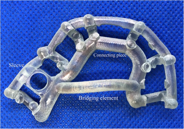
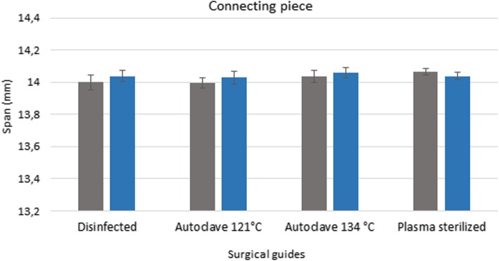
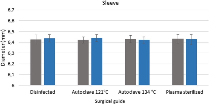
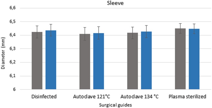
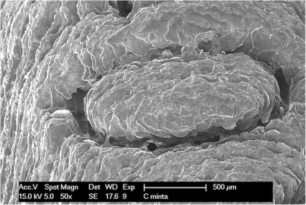
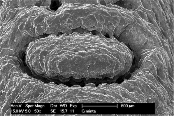
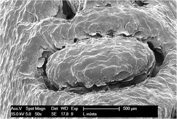
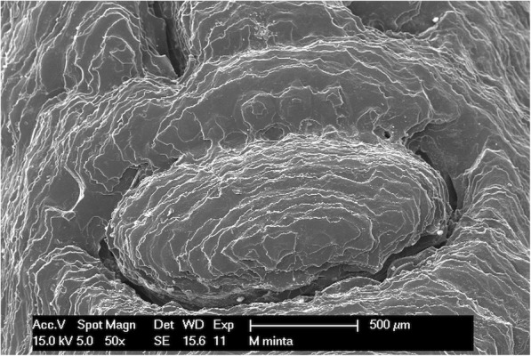
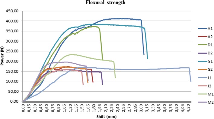
Fifteen identical drill guide templates were produced using a 3D printer. The surgical guides were classified into five groups: three controls, three disinfected (4% Gigasept®, 60 min), three plasma sterilized, three autoclave sterilized (+ 1 bar, 121 °C, 20 min), and three autoclave sterilized (+ 2 bar, 134 °C, 10 min). The templates were digitalized with a Steinbichler SCAN ST 3D scanner. Length was measured under an SZX16 stereomicroscope. A scanning electron microscope was used to study the surface morphology of the drill templates. The hardness, and flexural and compressive strength were measured to assess any changes in the physical characteristics of the material caused by sterilization. The drill guide templates were also examined with a Dage XiDAT 6600 X-ray. During the X-ray examinations, the following parameters were used: 100 kV voltage, 128 AVG averaging, 0.8 W power. One-way analysis of variance (ANOVA) was used to detect the difference between groups.
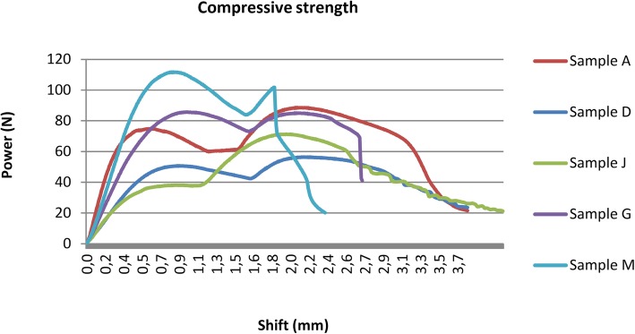
Evaluation of the hardness measurements of the various specimens shows that the hardness of the material was not changed by the plasma sterilization (p = 0.0680), steam sterilization on 121 °C (p = 0.6033) or disinfection process (p = 0.1399). The statistical analysis revealed significant difference in hardness strength of the autoclave sterilized (134 °C) specimens (p = 0.0002). There was no significant difference between the goups regarding the scanning electron microscopic and stereomicroscopic examinations. There was no significant difference regarding the X-ray visibility of the templates to the effect of the disinfection (p = 0.7844), plasma sterilization (p = 0.4091) and steam sterilization on 121 °C (p = 0.9277) and steam sterilization on 131 °C (p = 0.093). The effect of the sterilization was the same in case of both flexural and compressive strength of the material.
Our findings indicate that plasma sterilization and steam sterilization at 121 °C were both suitable for sterilizing the tested 3D printed surgical guides.
本研究旨在探讨消毒和三种不同的灭菌方法对用于种植体治疗的三维(3D)打印手术导板的尺寸变化和力学性能的影响。目的是使用破坏性和非破坏性的材料测试评估 3D 打印钻头导板模板的灭菌程序的效果。
使用 3D 打印机制作了 15 个相同的钻头导向模板。手术导板分为五组:三组对照,三组消毒(4% Gigasept®,60 分钟),三组等离子灭菌,三组高压灭菌锅灭菌(+1 巴,121°C,20 分钟),三组高压灭菌锅灭菌(+2 巴,134°C,10 分钟)。使用 Steinbichler SCAN ST 3D 扫描仪对模板进行数字化。在 SZX16 立体显微镜下测量长度。使用扫描电子显微镜研究钻头模板的表面形态。测量硬度、弯曲强度和压缩强度,以评估灭菌对材料物理特性的任何变化。还使用 Dage XiDAT 6600 X 射线对钻头导向模板进行了检查。在 X 射线检查中,使用了以下参数:100kV 电压、128AVG 平均、0.8W 功率。使用单向方差分析(ANOVA)来检测组间差异。
对各种样本的硬度测量评估表明,等离子灭菌(p=0.0680)、121°C 蒸汽灭菌(p=0.6033)或消毒过程(p=0.1399)均未改变材料的硬度。统计学分析显示,高压灭菌锅灭菌(134°C)标本的硬度强度有显著差异(p=0.0002)。在扫描电子显微镜和立体显微镜检查方面,各组之间没有显著差异。消毒(p=0.7844)、等离子灭菌(p=0.4091)和 121°C 蒸汽灭菌(p=0.9277)对模板 X 射线可见性以及 131°C 蒸汽灭菌(p=0.093)对模板 X 射线可见性没有显著差异。在材料的弯曲强度和压缩强度方面,灭菌的效果是相同的。
我们的研究结果表明,等离子灭菌和 121°C 蒸汽灭菌都适合于对测试的 3D 打印手术导板进行灭菌。