Department of Radiology, Okayama University Medical School, 2-5-1 Shikatacho, Kitaku, Okayama, 700-8558, Japan.
Intelligent Orthopaedic System Development, Okayama University Medical School, 2-5-1 Shikatacho, Kitaku, Okayama, 700-8558, Japan.
Eur Radiol Exp. 2020 Jan 28;4(1):4. doi: 10.1186/s41747-019-0135-0.
To reveal trends in bone microarchitectural parameters with increasing spatial resolution on ultra-high-resolution computed tomography (UHRCT) in vivo and to compare its performance with that of conventional-resolution CT (CRCT) and micro-CT ex vivo.
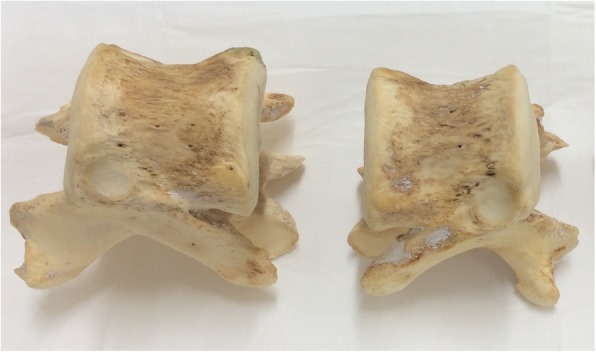
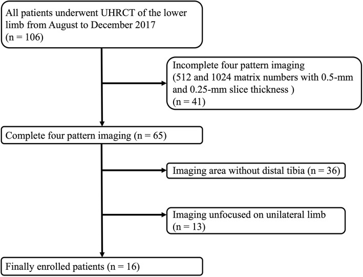
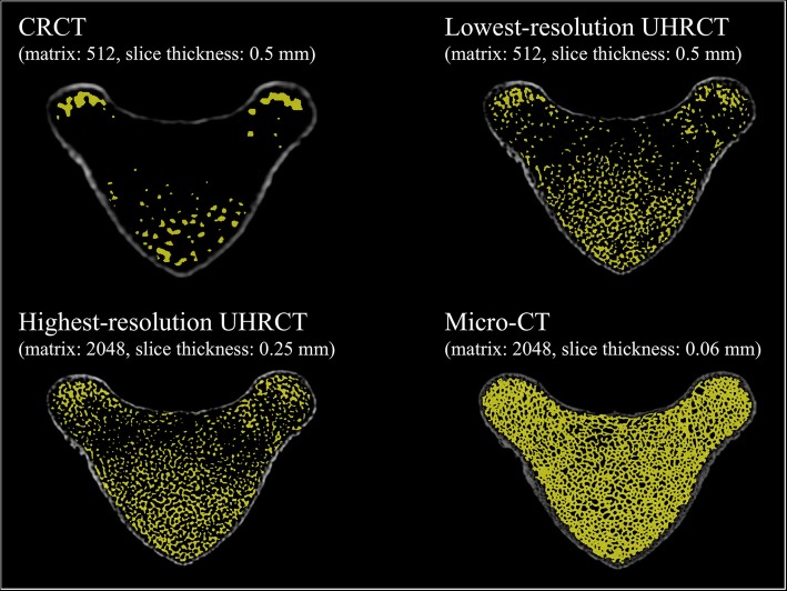
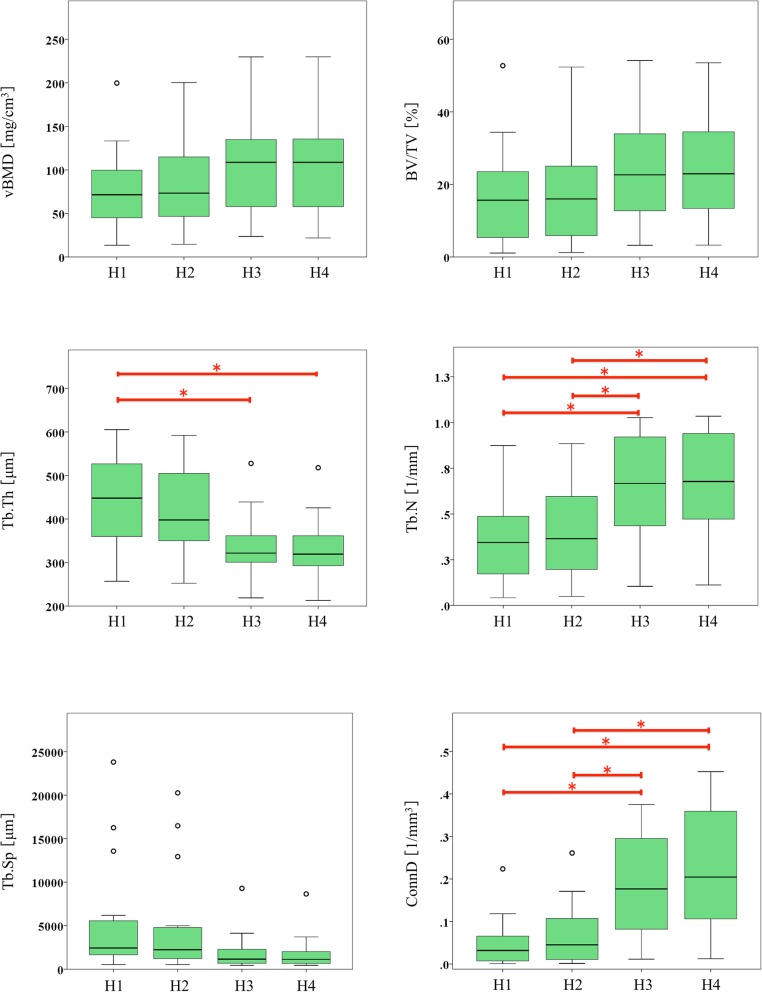
We retrospectively assessed 5 tiger vertebrae ex vivo and 16 human tibiae in vivo. Seven-pattern and four-pattern resolution imaging were performed on tiger vertebra using CRCT, UHRCT, and micro-CT, and on human tibiae using UHRCT. We measured six microarchitectural parameters: volumetric bone mineral density (vBMD), trabecular bone volume fraction (bone volume/total volume, BV/TV), trabecular thickness (Tb.Th), trabecular number (Tb.N), trabecular separation (Tb.Sp), and connectivity density (ConnD). Comparisons between different imaging resolutions were performed using Tukey or Dunnett T3 test.
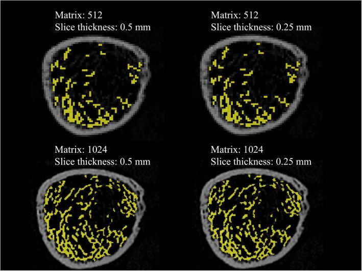
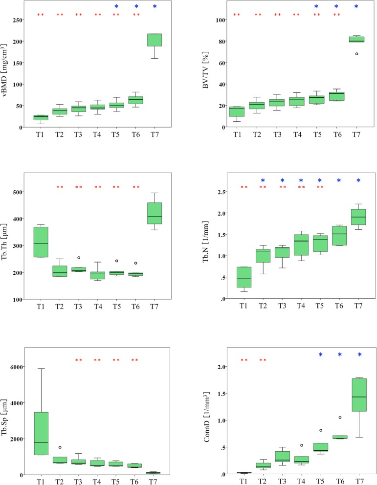
The vBMD, BV/TV, Tb.N, and ConnD parameters showed an increasing trend, while Tb.Sp showed a decreasing trend both ex vivo and in vivo. Ex vivo, UHRCT at the two highest resolutions (1024- and 2048-matrix imaging with 0.25-mm slice thickness) and CRCT showed significant differences (p ≤ 0.047) in vBMD (51.4 mg/cm and 63.5 mg/cm versus 20.8 mg/cm), BV/TV (26.5% and 29.5% versus 13.8 %), Tb.N (1.3 l/mm and 1.48 l/mm versus 0.47 l/mm), and ConnD (0.52 l/mm and 0.74 l/mm versus 0.02 l/mm, respectively). In vivo, the 512- and 1024-matrix imaging with 0.25-mm slice thickness showed significant differences in Tb.N (0.38 l/mm versus 0.67 l/mm, respectively) and ConnD (0.06 l/mm versus 0.22 l/mm, respectively).
We observed characteristic trends in microarchitectural parameters and demonstrated the potential utility of applying UHRCT for microarchitectural analysis.
揭示超高分辨率计算机断层扫描(UHRCT)体内骨微观结构参数随空间分辨率增加的趋势,并比较其与常规分辨率 CT(CRCT)和微 CT 体外的性能。
我们回顾性评估了 5 只老虎的 16 个人类胫骨。使用 CRCT、UHRCT 和微 CT 对老虎的 7 种和 4 种模式分辨率成像,以及对人类胫骨的 UHRCT 进行成像。我们测量了 6 个微观结构参数:体积骨矿物质密度(vBMD)、骨小梁体积分数(骨体积/总体积,BV/TV)、骨小梁厚度(Tb.Th)、骨小梁数量(Tb.N)、骨小梁间距(Tb.Sp)和连通密度(ConnD)。使用 Tukey 或 Dunnett T3 检验比较不同成像分辨率之间的差异。
vBMD、BV/TV、Tb.N 和 ConnD 参数在体内和体外均呈增加趋势,而 Tb.Sp 呈减少趋势。在体外,UHRCT 在两个最高分辨率(1024-和 2048-矩阵成像,层厚 0.25-mm)和 CRCT 之间显示出显著差异(p≤0.047),vBMD(51.4 mg/cm 和 63.5 mg/cm 与 20.8 mg/cm)、BV/TV(26.5%和 29.5%与 13.8%)、Tb.N(1.3 l/mm 和 1.48 l/mm 与 0.47 l/mm)和 ConnD(0.52 l/mm 和 0.74 l/mm 与 0.02 l/mm)。在体内,512-和 1024-矩阵成像,层厚 0.25-mm 显示 Tb.N(0.38 l/mm 与 0.67 l/mm)和 ConnD(0.06 l/mm 与 0.22 l/mm)的显著差异。
我们观察到微观结构参数的特征趋势,并证明了应用 UHRCT 进行微观结构分析的潜力。