Mangouritsas G, Koutropoulou N, Ragkousis A, Boutouri E, Diagourtas A
Eye Clinic, General Hospital ''Red Cross'', Athens, Greece.
1st University Eye Clinic, General Hospital "G. Gennimatas", Athens, Greece.
Clin Ophthalmol. 2019 Dec 13;13:2511-2519. doi: 10.2147/OPTH.S224757. eCollection 2019.
To investigate vessel density (VD) of radial peripapillary capillaries (RPC) and structural alterations in patients with unilateral preperimetric glaucoma (PPG) using optical coherence tomography angiography (OCTA).
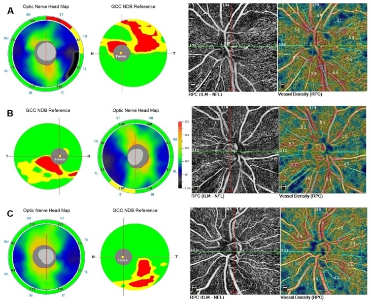
This cross-sectional observational study included 13 untreated patients with unilateral PPG. PPG eyes had larger excavation and abnormal thinning of retinal nerve fiber layer (RNFL) and/or ganglion cell complex (GCC) compared with fellow eyes (F). Both RNFL and GCC thickness in F were statistically within normal limits and/or borderline. The RPC VD on optic disc (idVD), of peripapillary (ppVD) and whole image (wiVD) scan area was measured. Twenty healthy eyes (H) served as controls. Structural and vascular parameters obtained by spectral-domain OCT/OCTA (Optovue; Fremont, CA) were compared between PPG, F and H.
Mean RNFL and GCC average thickness in microns differed significantly (p<0.001) between PPG (82.4±7.1, 81.4±5.9), F (91.0±7.1, 88.5±3.8) and H (103.5±6.0, 99.3±5.7). PPG compared with F showed significantly (p<0.001) lower mean ppVD (43.8%±3.0% versus 47.8%±3.2%) and wiVD (45.9%±3.5% versus 50.1%±3.9%). Mean ppVD (49.7%±2.4%) and wiVD (52.6%±3.0%) in H were not significantly higher than in F. Mean idVD showed no significant differences among the 3 groups. Areas under the receiver operating characteristic curves (AUROCs) for RNFL, GCC, ppVD and wiVD between PPG and H were excellent (>0.9). AUROCs between F and H demonstrated an excellent diagnostic ability for structural parameters and a poor one (<0.7) for vascular parameters.
Affected eyes of patients with unilateral PPG demonstrated significant RPC dropout. Clinically unaffected eyes showed thinner structural parameters but no significant microvasculature differences compared with non-glaucomatous eyes. Diagnostic ability of peripapillary vascular parameters was not superior to structural measurements. Microvascular dysfunction seems to be an early but not a primary event in glaucoma continuum at the stage of undetectable visual field loss. OCTA can be useful in early glaucoma diagnosis.
使用光学相干断层扫描血管造影(OCTA)研究单侧视野缺损前青光眼(PPG)患者的视盘周围径向毛细血管(RPC)血管密度(VD)及结构改变。
这项横断面观察性研究纳入了13例未经治疗的单侧PPG患者。与对侧眼(F)相比,PPG患眼的视盘凹陷更大,视网膜神经纤维层(RNFL)和/或神经节细胞复合体(GCC)异常变薄。F眼中RNFL和GCC厚度在统计学上均在正常范围内和/或临界值。测量视盘上(idVD)、视盘周围(ppVD)和全图像(wiVD)扫描区域的RPC VD。20只健康眼(H)作为对照。比较PPG组、F组和H组通过光谱域OCT/OCTA(Optovue;加利福尼亚州弗里蒙特)获得的结构和血管参数。
PPG组(82.4±7.1,81.4±5.9)、F组(91.0±7.1,88.5±3.8)和H组(103.5±6.0,99.3±5.7)的平均RNFL和GCC平均厚度(微米)差异有统计学意义(p<0.001)。与F组相比,PPG组的平均ppVD(43.8%±3.0%对47.8%±3.2%)和wiVD(45.9%±3.5%对50.1%±3.9%)显著更低(p<0.001)。H组的平均ppVD(49.7%±2.4%)和wiVD(52.6%±3.0%)并不显著高于F组。3组间的平均idVD无显著差异。PPG组和H组之间RNFL、GCC、ppVD和wiVD的受试者工作特征曲线下面积(AUROCs)极佳(>0.9)。F组和H组之间的AUROCs显示结构参数的诊断能力极佳,而血管参数的诊断能力较差(<0.7)。
单侧PPG患者的患眼表现出明显的RPC缺失。临床未受影响的眼睛显示结构参数较薄,但与非青光眼眼相比微血管无显著差异。视盘周围血管参数的诊断能力并不优于结构测量。在视野损失不可检测阶段,微血管功能障碍似乎是青光眼连续过程中的早期但非主要事件。OCTA在青光眼早期诊断中可能有用。