Pflüger Patrick, Pinnell Richard C, Martini Nadja, Hofmann Ulrich G
Section for Neuroelectronic Systems, Clinic for Neurosurgery, Medical Center - University of Freiburg, University of Freiburg, Freiburg im Breisgau, Germany.
Faculty of Medicine, University of Freiburg, Freiburg im Breisgau, Germany.
Front Neurosci. 2020 Jan 10;13:1367. doi: 10.3389/fnins.2019.01367. eCollection 2019.
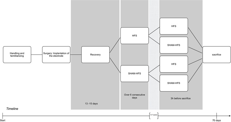
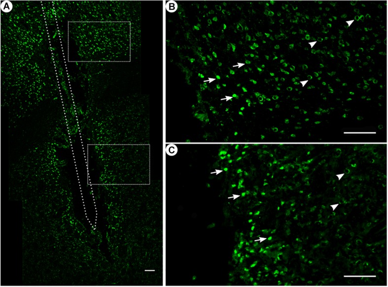
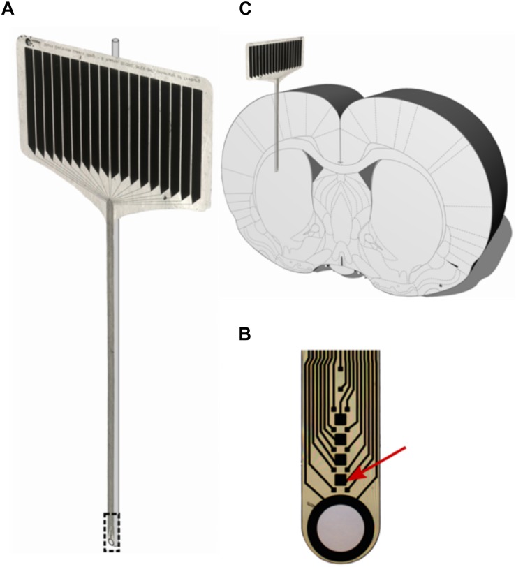
When designing electrodes and probes for brain-machine interfaces, one of the challenges faced involves minimizing the brain-tissue response, which would otherwise create an environment that is detrimental for the accurate functioning of such probes. Following the implantation process, the brain reacts with a sterile inflammation response and resulting astrocytic glial scar formation, potentially resulting in neuronal cell loss around the implantation site. Such alterations in the naïve brain tissue can hinder both the quality of neuronal recordings, and the efficacy of deep-brain stimulation. In this study, we chronically implanted a glass-supported polyimide microelectrode in the dorsolateral striatum of Sprague-Dawley rats. The effect of high-frequency stimulation (HFS) was investigated using immunoreactivity techniques. GFAP and ED1 immunohistochemistry were used to analyze the brain-tissue response. No changes in expression were found for either the acute or chronic stimulus groups; although a expression was found along the length of the implantation trajectory, following chronic implantation of our stiffened polyimide microelectrode. Furthermore, we also observed the formation of a glial scar around the microelectrode, with an accompanying low number of inflammation cells. Histological and statistical analysis of NeuN-positive cells did not demonstrate a pronounced "kill zone" with accompanying neuronal cell death around the implantation site, neither on the polymer side, nor on the glass side of the PI-glass probe.
在设计用于脑机接口的电极和探针时,面临的挑战之一是尽量减少脑组织反应,否则会营造出不利于此类探针准确发挥功能的环境。植入过程后,大脑会产生无菌性炎症反应并形成星形胶质细胞胶质瘢痕,这可能导致植入部位周围的神经元细胞丢失。幼稚脑组织的这种改变会妨碍神经元记录的质量以及深部脑刺激的效果。在本研究中,我们将玻璃支撑的聚酰亚胺微电极长期植入Sprague-Dawley大鼠的背外侧纹状体。使用免疫反应技术研究高频刺激(HFS)的效果。采用GFAP和ED1免疫组织化学分析脑组织反应。急性或慢性刺激组均未发现表达变化;尽管在我们的硬化聚酰亚胺微电极长期植入后,沿植入轨迹的长度发现了一种表达。此外,我们还观察到微电极周围形成了胶质瘢痕,伴有少量炎症细胞。对NeuN阳性细胞的组织学和统计分析未显示在植入部位周围聚合物侧或PI-玻璃探针的玻璃侧有明显的“杀伤区”以及伴随的神经元细胞死亡。