Department of Internal Medicine, Dankook University Hospital, Dankook University College of Medicine, Cheonan, Korea.
Department of Internal Medicine, Asan Medical Center, University of Ulsan College of Medicine, Seoul, Korea.
Gut Liver. 2020 Nov 15;14(6):826-832. doi: 10.5009/gnl19123.
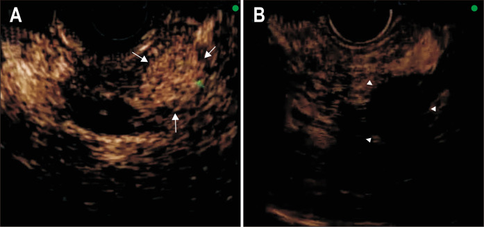
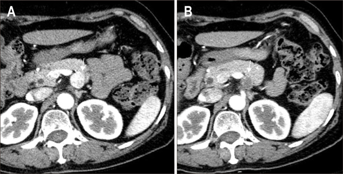
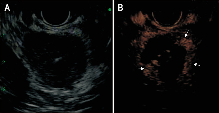
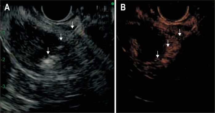
BACKGROUND/AIMS: Interventional endoscopists may utilize contrast-enhanced harmonic endoscopic ultrasound (CEHEUS) for image guidance during radiofrequency ablation (RFA) because of its capability to delineate real-time tumor perfusion dynamics. The purpose of this study was to assess the utility of CEH-EUS for the guidance and monitoring of endoscopic RFA.
Nineteen consecutive patients with solid abdominal tumors who underwent CEH-EUS and endoscopic RFA were included. The extent of the ablation was assessed by CEH-EUS at 5 to 7 days after RFA. Additional RFAs were performed under CEH-EUS guidance.
The diagnoses were as follows: nonfunctioning neuroendocrine tumor (n=13), solid pseudopapillary neoplasm (SPN) (n=2), insulinoma (n=1), left adrenal adenoma (n=2), and left adrenal oligometastasis (n=1). Pre-CEH-EUS findings revealed that 17 cases showed hyperenhanced patterns and two cases of SPN showed isoenhanced patterns. CEH-EUS-assisted RFA was technically feasible in all 19 patients. After the first RFA session, seven patients of the treated tumors showed the disappearance of intratumoral enhancement on CEH-EUS, whereas 12 showed residual contrast enhancement. Twelve patients with incomplete ablation were further treated with additional RFA under real-time CEH-EUS guidance. Radiologic complete response was observed in 13 patients (68.4%). Among the 35 ablation procedures, the only adverse events were two episodes of pancreatitis (5.7%; 1 moderate and 1 mild). During the median follow-up of 28 months, the local recurrence rate was 7.7%.
The application of CEH-EUS for RFA could be helpful in assessing early treatment response after ablation and targeting residual viable tumors during additional ablation sessions.
背景/目的:介入内镜医师可能会在射频消融 (RFA) 期间使用对比增强谐波内镜超声 (CEHEUS) 进行图像引导,因为它能够描绘实时肿瘤灌注动力学。本研究的目的是评估 CEH-EUS 在指导和监测内镜 RFA 中的效用。
19 例接受 CEH-EUS 和内镜 RFA 的连续实性腹部肿瘤患者被纳入研究。在 RFA 后 5 至 7 天,通过 CEH-EUS 评估消融范围。在 CEH-EUS 引导下进行额外的 RFA。
诊断结果如下:无功能神经内分泌肿瘤 (n=13)、实性假乳头状肿瘤 (SPN) (n=2)、胰岛素瘤 (n=1)、左肾上腺腺瘤 (n=2)和左肾上腺寡转移 (n=1)。CEH-EUS 前发现 17 例呈高增强模式,2 例 SPN 呈等增强模式。19 例患者均能成功进行 CEH-EUS 辅助 RFA。首次 RFA 治疗后,12 例肿瘤中有 7 例 CEH-EUS 显示肿瘤内增强消失,12 例肿瘤仍有对比增强。12 例不完全消融的患者在实时 CEH-EUS 引导下进一步接受额外的 RFA 治疗。13 例患者观察到放射学完全缓解 (68.4%)。在 35 次消融手术中,仅发生 2 例胰腺炎 (5.7%;1 例中度,1 例轻度)。在 28 个月的中位随访期间,局部复发率为 7.7%。
CEH-EUS 在 RFA 中的应用有助于评估消融后的早期治疗反应,并在额外的消融治疗中靶向残留的存活肿瘤。