Park Junha, Kim Taekwan, Kim Minah, Lee Tae Young, Kwon Jun Soo
Department of Medicine, Seoul National University College of Medicine, Seoul, Republic of Korea.
Department of Brain and Cognitive Sciences, Seoul National University College of Natural Sciences, Seoul, Republic of Korea.
Psychiatry Investig. 2020 Feb;17(2):87-95. doi: 10.30773/pi.2019.0206. Epub 2020 Feb 3.
It is well established that the cortico-striato-thalamo-cortical (CSTC) circuit is implicated in the pathophysiology of obsessive- compulsive disorder (OCD). However, reports on corticostriatal functional connectivity (FC) in OCD have been inconsistent due to the structural and functional heterogeneity of the striatum. Therefore, in the present study, we investigated corticostriatal FC using a fine 12-seed striatal parcellation to overcome this heterogeneity and discover the neural correlates of symptoms in OCD patients.
We recruited 23 OCD patients and 23 healthy controls (HCs). Whole-brain FC based on striatal seeds was examined using resting-state functional magnetic resonance imaging data and compared across OCD patients and HCs. We conducted correlation analysis between FCs of striatal subregions with significant group differences and symptom severity scores on the Yale-Brown Obsessive Compulsive Scale (Y-BOCS), Hamilton Rating Scale for Depression, and Hamilton Rating Scale for Anxiety (HAM-A).
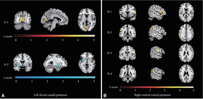
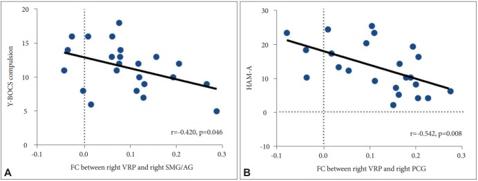
Compared to HCs, patients demonstrated increased FC of the dorsal caudal putamen and ventral rostral putamen (VRP) with several cortical regions, such as the intracalcarine cortex, inferior frontal gyrus, supramarginal/angular gyrus (SMG/AG), and postcentral gyrus (PCG). Furthermore, FC between the VRP and SMG/AG and between the VRP and PCG was negatively correlated with scores on the Y-BOCS compulsive subscale and the HAM-A, respectively.
These findings suggest that striatal subregions have strengthened FC with extensive cortical regions, which may reflect neural correlates of compulsive and anxious symptoms in OCD patients. These results contribute to an improved understanding of OCD pathophysiology by complementing the current evidence regarding striatal FC.
皮质-纹状体-丘脑-皮质(CSTC)回路与强迫症(OCD)的病理生理学有关,这一点已得到充分证实。然而,由于纹状体的结构和功能异质性,关于OCD中皮质纹状体功能连接(FC)的报道并不一致。因此,在本研究中,我们使用精细的12种子纹状体分区来研究皮质纹状体FC,以克服这种异质性,并发现OCD患者症状的神经相关性。
我们招募了23名OCD患者和23名健康对照(HCs)。使用静息态功能磁共振成像数据检查基于纹状体种子的全脑FC,并在OCD患者和HCs之间进行比较。我们对纹状体亚区域的FC与耶鲁-布朗强迫症量表(Y-BOCS)、汉密尔顿抑郁量表和汉密尔顿焦虑量表(HAM-A)上具有显著组间差异的症状严重程度评分进行了相关分析。
与HCs相比,患者表现出背侧尾状壳核和腹侧喙状壳核(VRP)与几个皮质区域(如距状沟内皮质、额下回、缘上回/角回(SMG/AG)和中央后回(PCG))之间的FC增加。此外,VRP与SMG/AG之间以及VRP与PCG之间的FC分别与Y-BOCS强迫分量表和HAM-A的评分呈负相关。
这些发现表明,纹状体亚区域与广泛的皮质区域之间的FC增强,这可能反映了OCD患者强迫和焦虑症状的神经相关性。这些结果通过补充当前关于纹状体FC的证据,有助于更好地理解OCD的病理生理学。