• 文献检索
  • 文档翻译
  • 深度研究
  • 学术资讯
  • Suppr Zotero 插件Zotero 插件
  • 邀请有礼
  • 套餐&价格
  • 历史记录
应用&插件
Suppr Zotero 插件Zotero 插件浏览器插件Mac 客户端Windows 客户端微信小程序
定价
高级版会员购买积分包购买API积分包
服务
文献检索文档翻译深度研究API 文档MCP 服务
关于我们
关于 Suppr公司介绍联系我们用户协议隐私条款
关注我们

Suppr 超能文献

核心技术专利:CN118964589B侵权必究
粤ICP备2023148730 号-1Suppr @ 2026

文献检索

告别复杂PubMed语法,用中文像聊天一样搜索,搜遍4000万医学文献。AI智能推荐,让科研检索更轻松。

立即免费搜索

文件翻译

保留排版,准确专业,支持PDF/Word/PPT等文件格式,支持 12+语言互译。

免费翻译文档

深度研究

AI帮你快速写综述,25分钟生成高质量综述,智能提取关键信息,辅助科研写作。

立即免费体验

细胞内ATP递送可导致组织快速再生,上调细胞因子、趋化因子和干细胞。

Intracellular ATP Delivery Causes Rapid Tissue Regeneration Upregulation of Cytokines, Chemokines, and Stem Cells.

作者信息

Mo Yiqun, Sarojini Harshini, Wan Rong, Zhang Qunwei, Wang Jianpu, Eichenberger Sarah, Kotwal Girish J, Chien Sufan

机构信息

Department of Environmental and Occupational Health Sciences, School of Public Health and Information Sciences, Louisville, KY, United States.

Department of Surgery, School of Medicine, University of Louisville, Louisville, KY, United States.

出版信息

Front Pharmacol. 2020 Jan 16;10:1502. doi: 10.3389/fphar.2019.01502. eCollection 2019.

DOI:10.3389/fphar.2019.01502
PMID:32009945
原文链接:https://pmc.ncbi.nlm.nih.gov/articles/PMC6976531/
Abstract

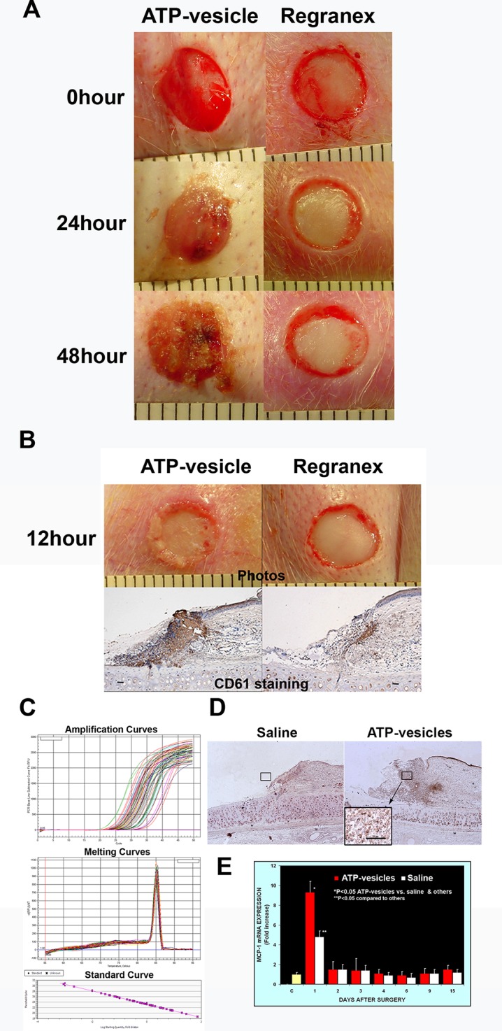
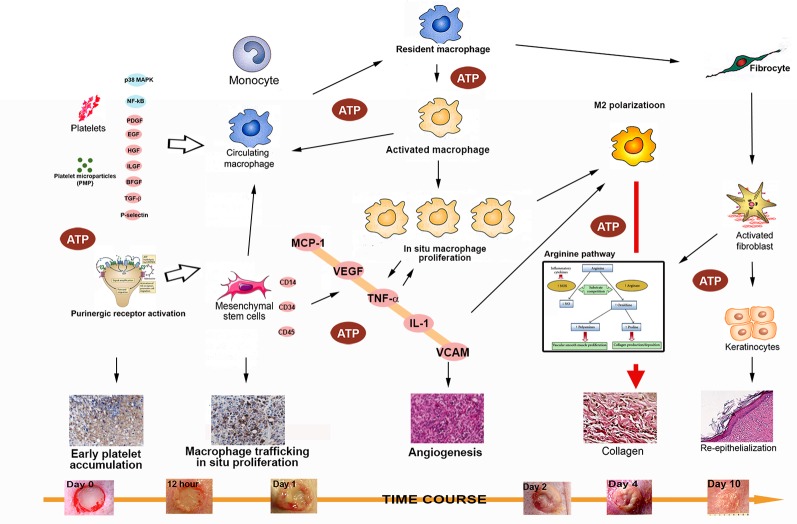
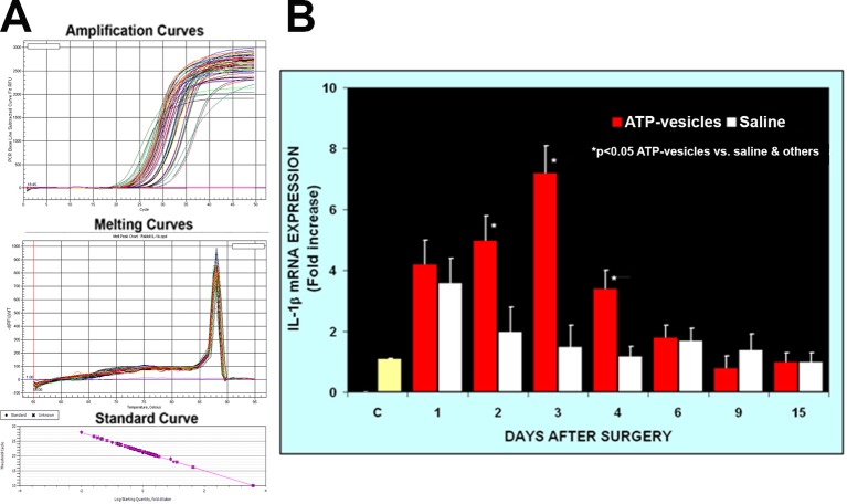
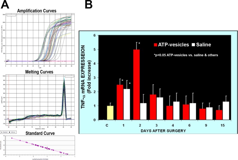
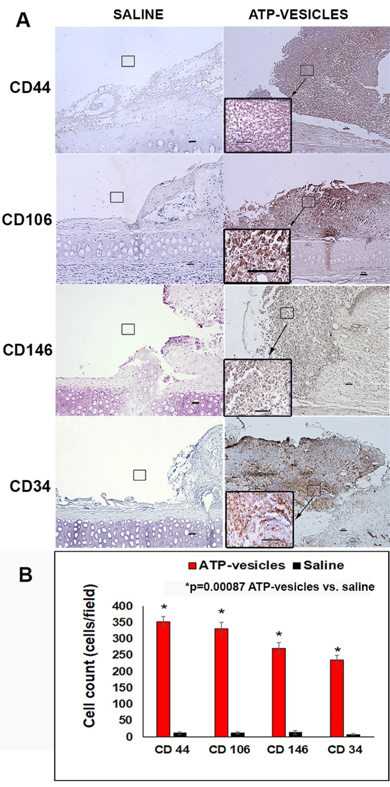
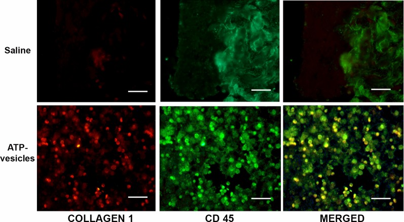
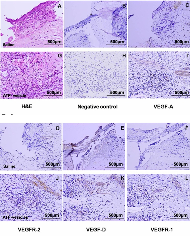
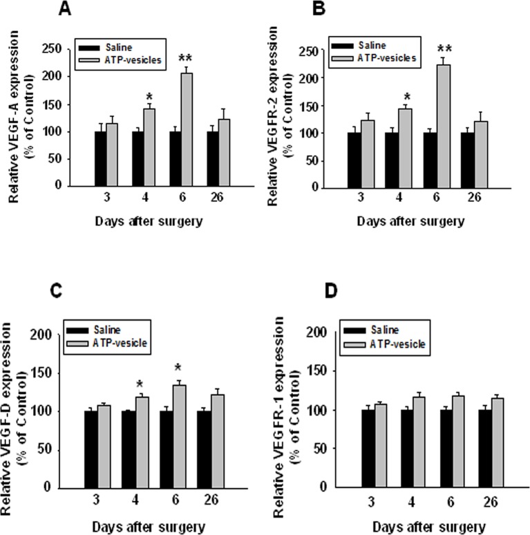
We have reported accelerated wound healing induced by intracellular ATP delivery in rabbits, through early massive accumulation, proliferation, and M2 polarization of macrophages. Granulation tissue started to grow within first 24 h of treatment and continued the growth till the wound cavity is completely covered. However, the mechanisms underlying this macrophage response are totally unclear because no one has ever reported this before. In this study, we performed a preliminary exploration of the possible mechanisms by focusing on the roles of cytokines, growth factors, and stem cells in this process. Among the 33 adult rabbits, 18 were used for cytokine measurements and the remaining were used for histological and immunohistochemical studies. Four wounds were created on the ventral side of each ear. Two wounds on one side were treated with ATP-vesicles (10 mM ATP), and the other two were treated with controls (normal saline or Regranex). Dressing changes were made daily and the rabbits were sacrificed at 5 h, 12 h, and 1, 2, 3, 4, 6, 9, 15, and 26 days after wounding. Tissue samples were analyzed for cytokines and growth factors using real-time PCR and immunohistochemical staining. The control wounds showed an immediate increase in proinflammatory cytokines after wound creation but no further increase after this initial spike. The growth factor levels in the control wounds remained unchanged throughout the study. Conversely, the wounds treated with ATP-vesicles showed significantly higher expression of MCP-1 and stem cell markers (CD44, CD106, CD146, and CD34) at day 1, significantly higher IL-1β and TNF-α expression from day 1-4, and significantly higher VEGF-A, VEGF-D, and VEGFR-2 expression from day 4-6 when compared to the controls. The significant upregulation of these factors corresponded to the very early and rapid macrophage accumulation, proliferation, and M2 polarization, resulting in unprecedented rapid granulation tissue generation due to direct macrophage collagen production and neovascularization.

摘要

我们曾报道,通过巨噬细胞的早期大量聚集、增殖和M2极化,细胞内ATP递送可加速家兔伤口愈合。肉芽组织在治疗的最初24小时内开始生长,并持续生长直至伤口腔完全被覆盖。然而,这种巨噬细胞反应的潜在机制完全不清楚,因为此前从未有人报道过。在本研究中,我们通过关注细胞因子、生长因子和干细胞在此过程中的作用,对可能的机制进行了初步探索。在33只成年家兔中,18只用于细胞因子测量,其余用于组织学和免疫组织化学研究。在每只耳朵的腹侧制造4个伤口。一侧的2个伤口用ATP囊泡(10 mM ATP)治疗,另外2个用对照(生理盐水或Regranex)治疗。每天更换敷料,并在受伤后5小时、12小时以及1、2、3、4、6、9、15和26天处死家兔。使用实时PCR和免疫组织化学染色分析组织样本中的细胞因子和生长因子。对照伤口在伤口形成后促炎细胞因子立即增加,但在最初的峰值后没有进一步增加。在整个研究过程中,对照伤口中的生长因子水平保持不变。相反,与对照相比,用ATP囊泡治疗的伤口在第1天显示MCP-1和干细胞标志物(CD44、CD106、CD146和CD34)的表达显著更高,在第1 - 4天IL-1β和TNF-α表达显著更高,在第4 - 6天VEGF-A、VEGF-D和VEGFR-2表达显著更高。这些因子的显著上调与巨噬细胞非常早期和快速的聚集、增殖和M2极化相对应,由于巨噬细胞直接产生胶原蛋白和血管生成,导致前所未有的快速肉芽组织生成。

https://cdn.ncbi.nlm.nih.gov/pmc/blobs/4912/6976531/bffeab43540f/fphar-10-01502-g008.jpg
https://cdn.ncbi.nlm.nih.gov/pmc/blobs/4912/6976531/80222543d89e/fphar-10-01502-g001.jpg
https://cdn.ncbi.nlm.nih.gov/pmc/blobs/4912/6976531/5327325eea0a/fphar-10-01502-g002.jpg
https://cdn.ncbi.nlm.nih.gov/pmc/blobs/4912/6976531/fc46a957979e/fphar-10-01502-g003.jpg
https://cdn.ncbi.nlm.nih.gov/pmc/blobs/4912/6976531/44406005826d/fphar-10-01502-g004.jpg
https://cdn.ncbi.nlm.nih.gov/pmc/blobs/4912/6976531/b6d9a3a7a411/fphar-10-01502-g005.jpg
https://cdn.ncbi.nlm.nih.gov/pmc/blobs/4912/6976531/bf23dd770d44/fphar-10-01502-g006.jpg
https://cdn.ncbi.nlm.nih.gov/pmc/blobs/4912/6976531/34f6d2bb99a1/fphar-10-01502-g007.jpg
https://cdn.ncbi.nlm.nih.gov/pmc/blobs/4912/6976531/bffeab43540f/fphar-10-01502-g008.jpg

相似文献

1
Intracellular ATP Delivery Causes Rapid Tissue Regeneration Upregulation of Cytokines, Chemokines, and Stem Cells.细胞内ATP递送可导致组织快速再生,上调细胞因子、趋化因子和干细胞。
Front Pharmacol. 2020 Jan 16;10:1502. doi: 10.3389/fphar.2019.01502. eCollection 2019.
2
Rapid granulation tissue regeneration by intracellular ATP delivery--a comparison with Regranex.通过细胞内递送ATP实现快速肉芽组织再生——与Regranex的比较
PLoS One. 2014 Mar 17;9(3):e91787. doi: 10.1371/journal.pone.0091787. eCollection 2014.
3
Rapid tissue regeneration induced by intracellular ATP delivery-A preliminary mechanistic study.细胞内递送ATP诱导的快速组织再生——一项初步机制研究。
PLoS One. 2017 Apr 5;12(4):e0174899. doi: 10.1371/journal.pone.0174899. eCollection 2017.
4
Macrophage Differentiation in Normal and Accelerated Wound Healing.正常和加速伤口愈合过程中的巨噬细胞分化
Results Probl Cell Differ. 2017;62:353-364. doi: 10.1007/978-3-319-54090-0_14.
5
Enhanced Skin Incisional Wound Healing With Intracellular ATP Delivery Macrophage Proliferation and Direct Collagen Production.通过细胞内ATP递送增强皮肤切口伤口愈合:巨噬细胞增殖和直接胶原蛋白生成
Front Pharmacol. 2021 Jun 16;12:594586. doi: 10.3389/fphar.2021.594586. eCollection 2021.
6
Intracellular adenosine triphosphate delivery enhanced skin wound healing in rabbits.细胞内三磷酸腺苷递送促进兔皮肤伤口愈合。
Ann Plast Surg. 2009 Feb;62(2):180-6. doi: 10.1097/SAP.0b013e31817fe47e.
7
[Effects and mechanism of rat epidermal stem cells treated with exogenous vascular endothelial growth factor on healing of deep partial-thickness burn wounds in rats].外源性血管内皮生长因子处理的大鼠表皮干细胞对大鼠深Ⅱ度烧伤创面愈合的影响及机制
Zhonghua Shao Shang Za Zhi. 2020 Mar 20;36(3):195-203. doi: 10.3760/cma.j.cn501120-20191125-00441.
8
[Preliminary evaluation and mechanism of adipose-derived stem cell transplantation from allogenic diabetic rats in the treatment of diabetic rat wounds].[同种异体糖尿病大鼠脂肪间充质干细胞移植治疗糖尿病大鼠创面的初步评价及机制]
Zhonghua Shao Shang Za Zhi. 2019 Sep 20;35(9):645-654. doi: 10.3760/cma.j.issn.1009-2587.2019.09.002.
9
Pivotal role of ATP in macrophages fast tracking wound repair and regeneration.ATP在巨噬细胞快速追踪伤口修复和再生中的关键作用。
Wound Repair Regen. 2015 Sep;23(5):724-7. doi: 10.1111/wrr.12323. Epub 2015 Sep 14.
10
[Effects of basic fibroblast growth factor on healing of infective wound in New Zealand rabbit after debridement].[碱性成纤维细胞生长因子对新西兰兔清创后感染性伤口愈合的影响]
Zhonghua Shao Shang Za Zhi. 2019 Feb 20;35(2):95-103. doi: 10.3760/cma.j.issn.1009-2587.2019.02.004.

引用本文的文献

1
Purinergic receptors play a key role in shock wave-induced proliferation.嘌呤能受体在冲击波诱导的增殖中起关键作用。
Sci Rep. 2025 May 31;15(1):19138. doi: 10.1038/s41598-025-02955-3.
2
Vanillin Enhances Photobiomodulation Wound Healing by Modulating Glyco-Oxidative Stress and Glucose Dysmetabolism in Diabetic Wounded Fibroblast Cells.香草醛通过调节糖尿病创伤成纤维细胞中的糖氧化应激和葡萄糖代谢异常来增强光生物调节伤口愈合。
J Cell Mol Med. 2025 Apr;29(7):e70537. doi: 10.1111/jcmm.70537.
3
Hyperbaric oxygen therapy in the ATLS/ACLS resuscitative management of acutely ill or severely injured patients with severe anemia: a review.

本文引用的文献

1
Significant upregulation of U1 and U4 spliceosomal snRNAs by ATP nanoliposomes explains acceleration of wound healing, due to increased pre-mRNA processing to functional mRNA.ATP 纳米脂质体显著上调 U1 和 U4 剪接体 snRNA,从而加速伤口愈合,这是由于前体 mRNA 加工为功能性 mRNA 的增加。
Nanomedicine. 2018 Jun;14(4):1289-1299. doi: 10.1016/j.nano.2018.03.003. Epub 2018 Apr 5.
2
Rapid tissue regeneration induced by intracellular ATP delivery-A preliminary mechanistic study.细胞内递送ATP诱导的快速组织再生——一项初步机制研究。
PLoS One. 2017 Apr 5;12(4):e0174899. doi: 10.1371/journal.pone.0174899. eCollection 2017.
3
高压氧疗法在严重贫血的急重症或重伤患者的ATLS/ACLS复苏管理中的应用:一项综述
Front Med (Lausanne). 2024 Oct 8;11:1408816. doi: 10.3389/fmed.2024.1408816. eCollection 2024.
4
Intercellular Communication in Airway Epithelial Cell Regeneration: Potential Roles of Connexins and Pannexins.细胞间通讯在气道上皮细胞再生中的作用:连接蛋白和pannexin 的潜在作用。
Int J Mol Sci. 2023 Nov 10;24(22):16160. doi: 10.3390/ijms242216160.
5
Mitochondria as a therapeutic: a potential new frontier in driving the shift from tissue repair to regeneration.线粒体作为一种治疗手段:推动从组织修复向再生转变的潜在新领域。
Regen Biomater. 2023 Aug 12;10:rbad070. doi: 10.1093/rb/rbad070. eCollection 2023.
6
Roles of extracellular adenosine triphosphate on the functions of periodontal ligament cells.细胞外三磷酸腺苷对牙周膜细胞功能的作用。
BDJ Open. 2023 Jul 8;9(1):28. doi: 10.1038/s41405-023-00147-7.
7
Natural γδT17 cell development and functional acquisition is governed by the mTORC2--Maf-controlled mitochondrial fission pathway.天然γδT17细胞的发育和功能获得受mTORC2-Maf控制的线粒体分裂途径调控。
iScience. 2023 Apr 10;26(5):106630. doi: 10.1016/j.isci.2023.106630. eCollection 2023 May 19.
8
The role of cluster of differentiation 163-positive macrophages in wound healing: a preliminary study and a systematic review.分化簇163阳性巨噬细胞在伤口愈合中的作用:一项初步研究及系统评价
Arch Dermatol Res. 2023 Apr;315(3):359-370. doi: 10.1007/s00403-022-02407-2. Epub 2022 Oct 25.
9
ENERGI-F703 gel, as a new topical treatment for diabetic foot and leg ulcers: A multicenter, randomized, double-blind, phase II trial.ENERGI-F703凝胶作为糖尿病足和腿部溃疡的一种新型局部治疗方法:一项多中心、随机、双盲、II期试验。
EClinicalMedicine. 2022 Jul 10;51:101497. doi: 10.1016/j.eclinm.2022.101497. eCollection 2022 Sep.
10
Polyphosphate in Chronic Wound Healing: Restoration of Impaired Metabolic Energy State.多聚磷酸盐在慢性伤口愈合中的作用:恢复受损的代谢能量状态。
Prog Mol Subcell Biol. 2022;61:51-82. doi: 10.1007/978-3-031-01237-2_4.
Mesenchymal stem cells: The roles and functions in cutaneous wound healing and tumor growth.
间充质干细胞:在皮肤伤口愈合和肿瘤生长中的作用与功能
J Dermatol Sci. 2017 May;86(2):83-89. doi: 10.1016/j.jdermsci.2016.11.005. Epub 2016 Nov 11.
4
Platelet Activation: The Mechanisms and Potential Biomarkers.血小板活化:机制与潜在生物标志物
Biomed Res Int. 2016;2016:9060143. doi: 10.1155/2016/9060143. Epub 2016 Jun 15.
5
Chemokine and chemokine receptors in autoimmunity: the case of primary biliary cholangitis.自身免疫中的趋化因子与趋化因子受体:原发性胆汁性胆管炎的实例
Expert Rev Clin Immunol. 2016 Jun;12(6):661-72. doi: 10.1586/1744666X.2016.1147956. Epub 2016 Feb 25.
6
Macrophage PPARγ and impaired wound healing in type 2 diabetes.巨噬细胞PPARγ与2型糖尿病患者伤口愈合受损
J Pathol. 2015 Aug;236(4):433-44. doi: 10.1002/path.4548. Epub 2015 May 12.
7
Rapid granulation tissue regeneration by intracellular ATP delivery--a comparison with Regranex.通过细胞内递送ATP实现快速肉芽组织再生——与Regranex的比较
PLoS One. 2014 Mar 17;9(3):e91787. doi: 10.1371/journal.pone.0091787. eCollection 2014.
8
MCP-1-induced protein promotes endothelial-like and angiogenic properties in human bone marrow monocytic cells.MCP-1 诱导蛋白促进人骨髓单核细胞的内皮样和血管生成特性。
J Pharmacol Exp Ther. 2013 Nov;347(2):288-97. doi: 10.1124/jpet.113.207316. Epub 2013 Sep 5.
9
Inflammation and wound healing: the role of the macrophage.炎症与伤口愈合:巨噬细胞的作用。
Expert Rev Mol Med. 2011 Jul 11;13:e23. doi: 10.1017/S1462399411001943.
10
Macrophage pro-inflammatory cytokine secretion is enhanced following interaction with autologous platelets.巨噬细胞与自身血小板相互作用后,促炎细胞因子的分泌增强。
J Inflamm (Lond). 2010 Nov 11;7:53. doi: 10.1186/1476-9255-7-53.