Centre for the Endothelium, Vascular Biology Program, Centenary Institute, The University of Sydney, Locked Bag 6, Newtown, Sydney, 2042, Australia.
Cell Commun Signal. 2020 Feb 3;18(1):18. doi: 10.1186/s12964-020-0511-7.
Vascular endothelial cell alignment in the direction of flow is an adaptive response that protects against aortic diseases such as atherosclerosis. The RhoGTPases are known to regulate this alignment. We have shown previously that ARHGAP18 in endothelial cells is a negative regulator of RhoC and its expression is essential in flow-mediated alignment. Depletion of ARHGAP18 inhibits alignment and results in the induction of a pro-inflammatory phenotype. In embryogenesis, ARHGAP18 was identified as a downstream effector of the Yes-associated protein, YAP, which regulates cell shape and size.
We have used siRNA technology to deplete either ARHGAP18 or YAP in human endothelial cells. The in vitro studies were performed under athero-protective, laminar flow conditions. The analysis of YAP activity was also investigated, using high performance confocal imaging, in our ARHGAP18 knockout mutant mice.
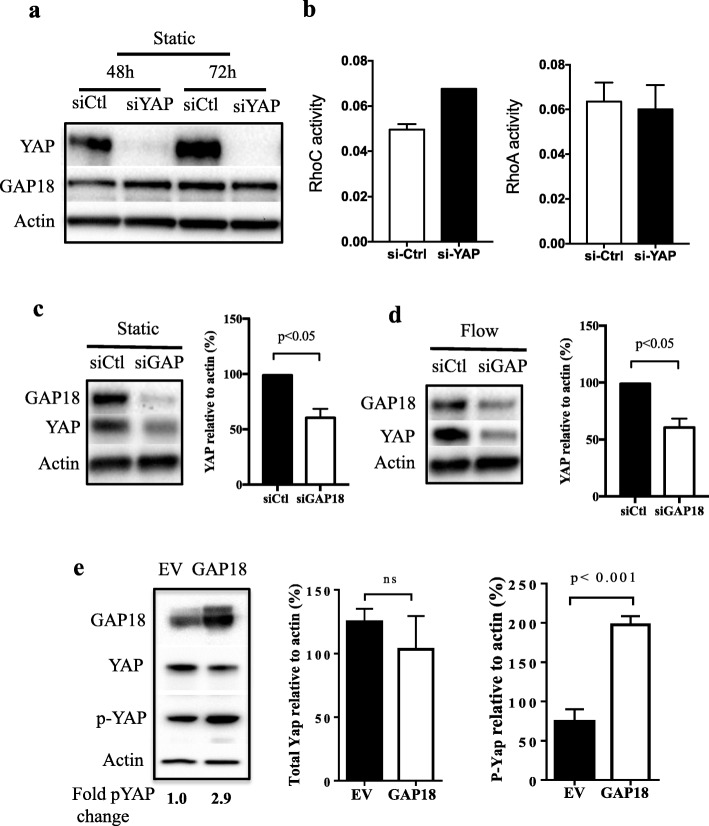
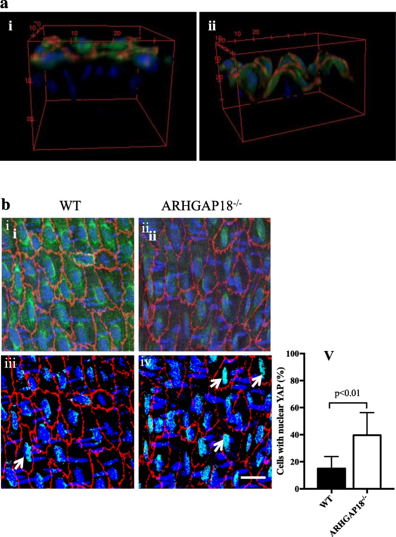
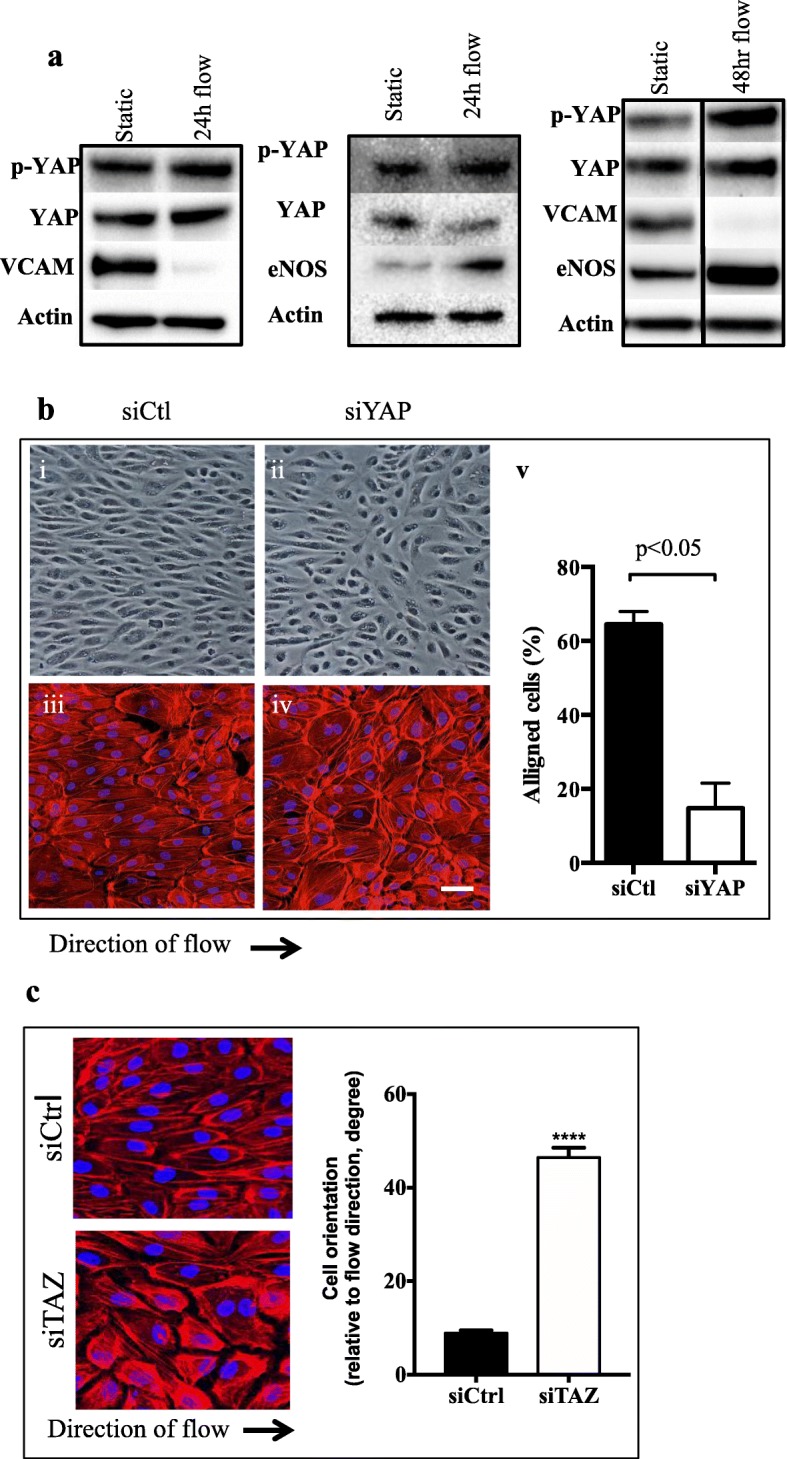
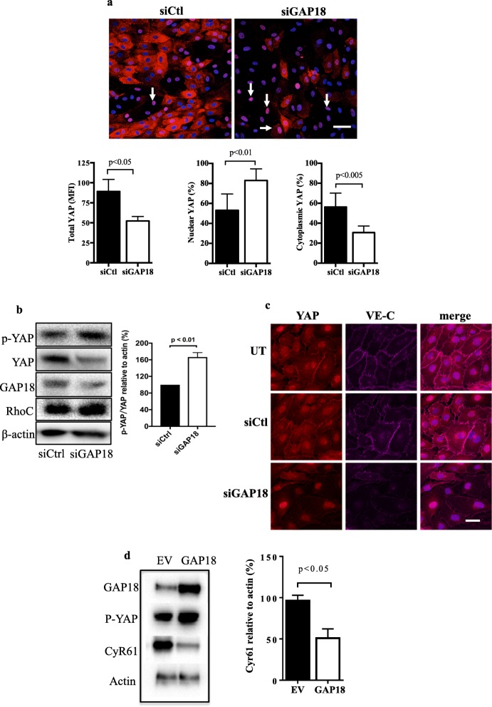
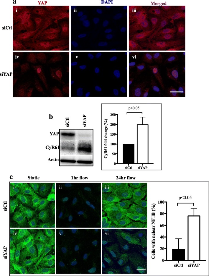
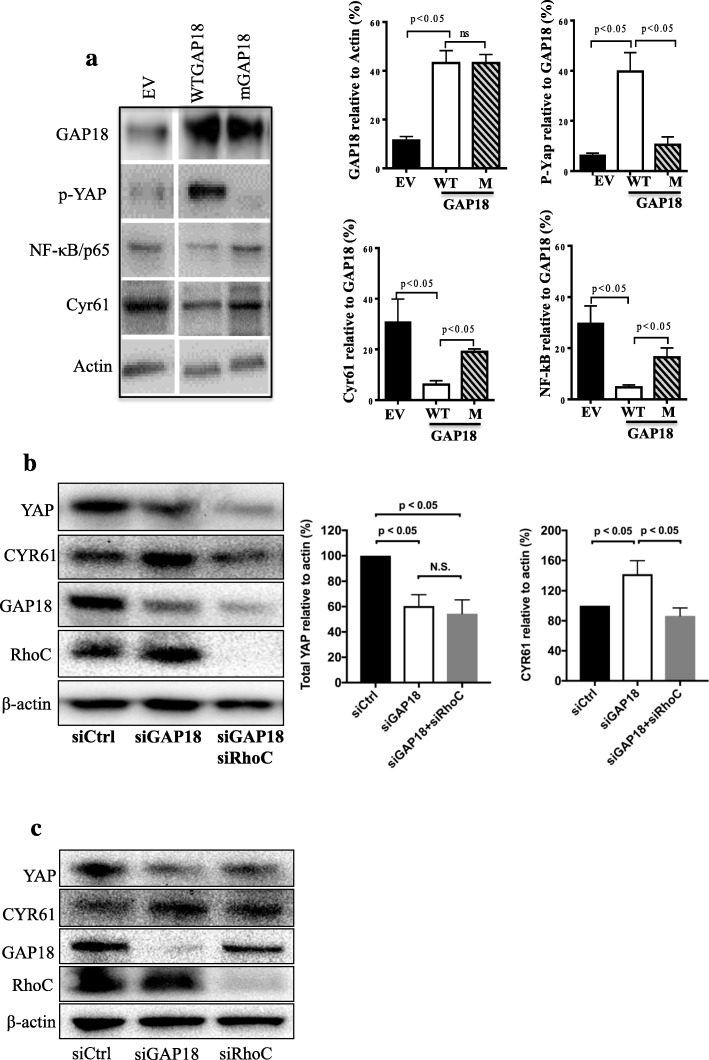
We show here that loss of ARHGAP18, although decreasing the expression of YAP results in its nuclear localisation consistent with activation. We further show that depletion of YAP itself results in its activation as defined by an in increase in its nuclear localisation and an increase in the YAP target gene, CyR61. Depletion of YAP, similar to that observed for ARHGAP18 depletion, results in loss of endothelial cell alignment under high shear stress mediated flow and also in the activation of NFkB, as determined by p65 nuclear localisation. In contrast, ARHGAP18 overexpression results in upregulation of YAP, its phosphorylation, and a decrease in the YAP target gene Cyr61, consistent with YAP inactivation. Finally, in ARHGAP18 deleted mice, in regions where there is a loss of endothelial cell alignment, a situation associated with a priming of the cells to a pro-inflammatory phenotype, YAP shows nuclear localisation.
Our results show that YAP is downstream of ARHGAP18 in mature endothelial cells and that this pathway is involved in the athero-protective alignment of endothelial cells under laminar shear stress. ARHGAP18 depletion leads to a disruption of the junctions as seen by loss of VE-Cadherin localisation to these regions and a concomitant localisation of YAP to the nucleus.
血管内皮细胞沿血流方向排列是一种适应性反应,可以预防主动脉疾病,如动脉粥样硬化。RhoGTPases 被认为可以调节这种排列。我们之前已经表明,内皮细胞中的 ARHGAP18 是 RhoC 的负调节剂,其表达对于血流介导的排列是必不可少的。ARHGAP18 的耗竭抑制排列并导致诱导促炎表型。在胚胎发生中,ARHGAP18 被鉴定为 Yes 相关蛋白 YAP 的下游效应物,YAP 调节细胞形状和大小。
我们使用 siRNA 技术耗尽人内皮细胞中的 ARHGAP18 或 YAP。在动脉保护、层流条件下进行体外研究。还使用高性能共聚焦成像分析了我们的 ARHGAP18 敲除突变小鼠中的 YAP 活性。
我们在这里表明,尽管 ARHGAP18 的缺失降低了 YAP 的表达,但导致其核定位一致,表明其激活。我们进一步表明,YAP 的耗竭本身会导致其激活,表现为核定位增加和 YAP 靶基因 CyR61 增加。与 ARHGAP18 耗竭观察到的情况一样,在高剪切力介导的流动下,YAP 的耗竭导致内皮细胞排列丧失,并且 NFkB 激活,如 p65 核定位所示。相比之下,ARHGAP18 的过表达导致 YAP 的上调、磷酸化和 YAP 靶基因 Cyr61 的减少,这与 YAP 失活一致。最后,在 ARHGAP18 缺失的小鼠中,在内皮细胞排列丧失的区域,即与细胞向促炎表型激活相关的区域,YAP 显示核定位。
我们的结果表明,YAP 是成熟内皮细胞中 ARHGAP18 的下游,该途径参与了层流剪切应力下内皮细胞的动脉保护排列。ARHGAP18 的耗竭导致细胞连接的破坏,如 VE-Cadherin 定位到这些区域的缺失以及 YAP 向核的共定位所见。