Bartolotta Tommaso Vincenzo, Terranova Maria Chiara, Gagliardo Cesare, Taibbi Adele
BiND Department: Biomedicine, Neuroscience and Advanced Diagnostic, University of Palermo, Via Del Vespro, 129 90127, Palermo, Italy.
Department of Radiology, Fondazione Istituto Giuseppe Giglio Ct.da Pietrapollastra, Via Pisciotto, 90015, Cefalù (Palermo), Italy.
Insights Imaging. 2020 Feb 4;11(1):9. doi: 10.1186/s13244-019-0819-2.
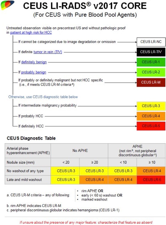
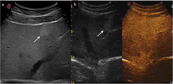
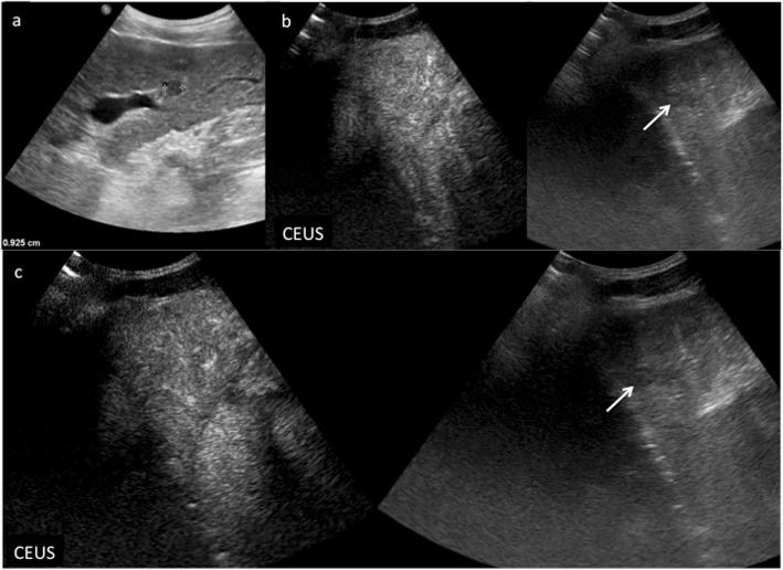
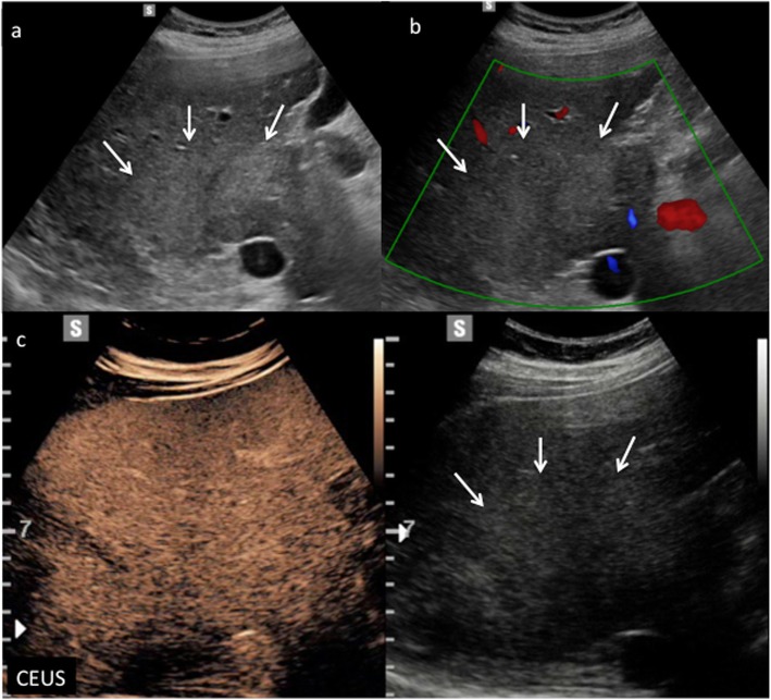
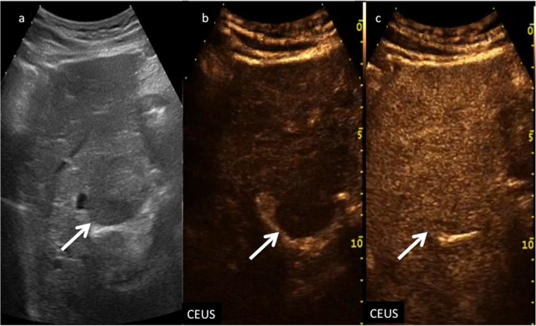
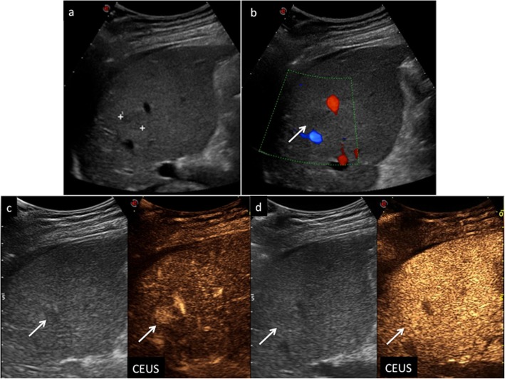
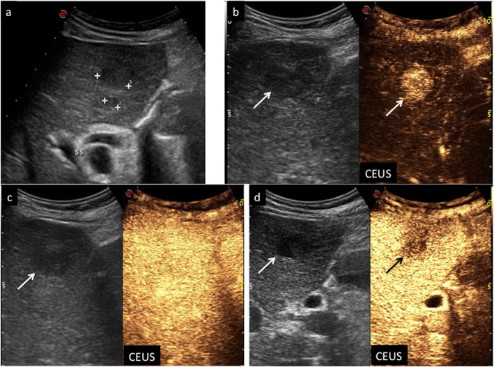
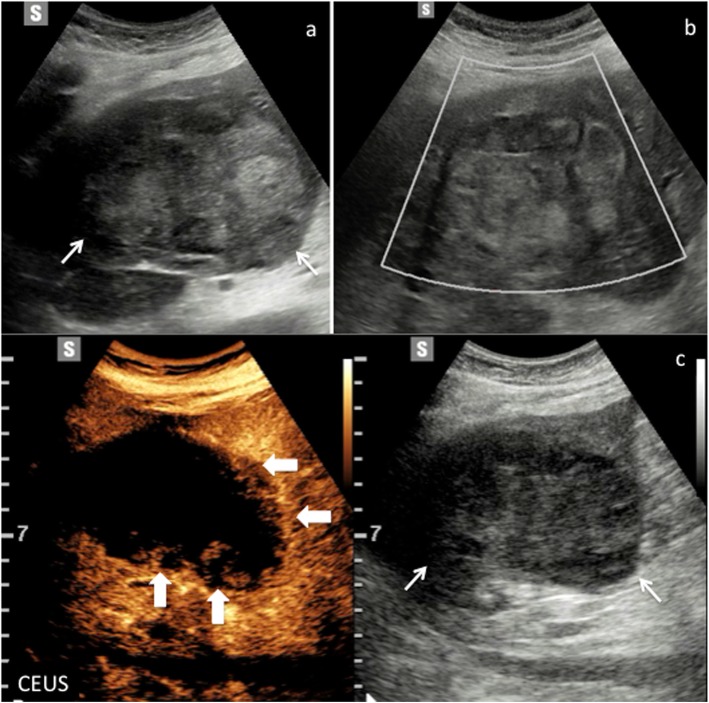
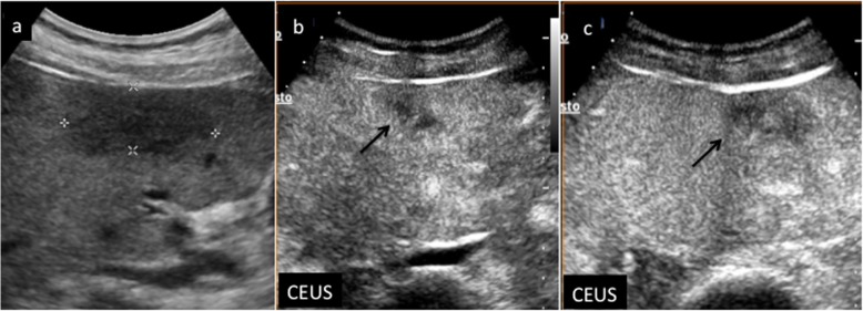
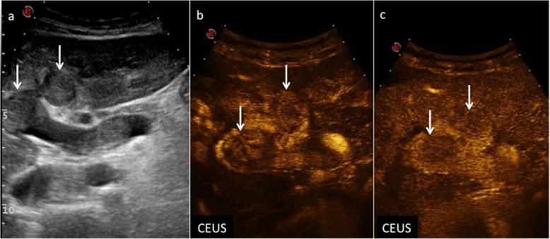
Contrast-enhanced ultrasound (CEUS) greatly improved the diagnostic accuracy of US in the detection and characterization of focal liver lesions (FLLs), and it is suggested and often included in many international guidelines as an important diagnostic tool in the imaging work-up of cirrhotic patients at risk for developing hepatocellular carcinoma (HCC). In particular, CEUS Liver Imaging Reporting and Data System (LI-RADS) provides standardized terminology, interpretation, and reporting for the diagnosis of HCC. The aim of this pictorial essay is to illustrate CEUS features of nodules discovered at US in cirrhotic liver according to LI-RADS categorization.
超声造影(CEUS)极大地提高了超声在检测和鉴别肝脏局灶性病变(FLLs)方面的诊断准确性,并且在许多国际指南中被推荐并经常作为对有肝细胞癌(HCC)发生风险的肝硬化患者进行影像学检查的重要诊断工具。特别是,CEUS肝脏影像报告和数据系统(LI-RADS)为HCC的诊断提供了标准化的术语、解读和报告。本图谱文章的目的是根据LI-RADS分类阐述在肝硬化肝脏超声检查中发现的结节的CEUS特征。