• 文献检索
  • 文档翻译
  • 深度研究
  • 学术资讯
  • Suppr Zotero 插件Zotero 插件
  • 邀请有礼
  • 套餐&价格
  • 历史记录
应用&插件
Suppr Zotero 插件Zotero 插件浏览器插件Mac 客户端Windows 客户端微信小程序
定价
高级版会员购买积分包购买API积分包
服务
文献检索文档翻译深度研究API 文档MCP 服务
关于我们
关于 Suppr公司介绍联系我们用户协议隐私条款
关注我们

Suppr 超能文献

核心技术专利:CN118964589B侵权必究
粤ICP备2023148730 号-1Suppr @ 2026

文献检索

告别复杂PubMed语法,用中文像聊天一样搜索,搜遍4000万医学文献。AI智能推荐,让科研检索更轻松。

立即免费搜索

文件翻译

保留排版,准确专业,支持PDF/Word/PPT等文件格式,支持 12+语言互译。

免费翻译文档

深度研究

AI帮你快速写综述,25分钟生成高质量综述,智能提取关键信息,辅助科研写作。

立即免费体验

大鼠眼内压升高对三层视网膜血管网络的反应。

Response of the Trilaminar Retinal Vessel Network to Intraocular Pressure Elevation in Rat Eyes.

机构信息

,.

出版信息

Invest Ophthalmol Vis Sci. 2020 Feb 7;61(2):2. doi: 10.1167/iovs.61.2.2.

DOI:10.1167/iovs.61.2.2
PMID:32031574
原文链接:https://pmc.ncbi.nlm.nih.gov/articles/PMC7325622/
Abstract

PURPOSE

The purpose of this study was to test the hypothesis that the superficial, intermediate, and deep retinal vascular plexus show different responses to intraocular pressure (IOP) elevation.

METHODS

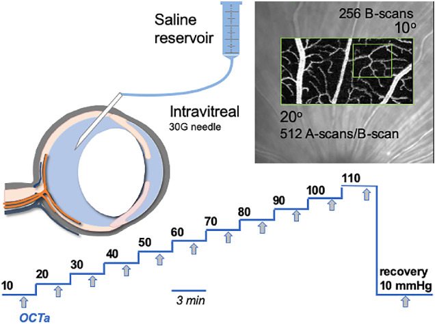
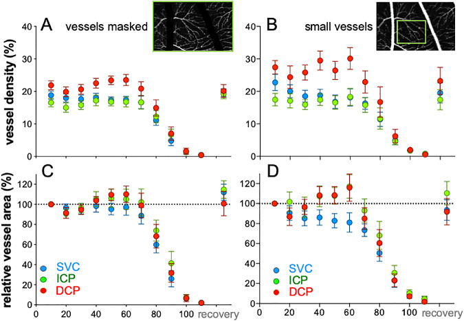
Anesthetized adult Long Evans rats (n = 14) were imaged using optical coherence tomography angiography (OCTA; Spectralis) at baseline (IOP 10 mm Hg) and in follow-up mode to examine the vasculature during IOP elevation (10 to 110 mm Hg, 10 mm Hg steps, each step 3 minutes). A 20° × 10° field was imaged. Vessel density within a 2D projection image was determined (%) for the superficial vascular complex (SVC), intermediate capillary plexus (ICP), and deep capillary plexus (DCP). Comparisons were made between layers using 2-way repeated measures ANOVA (layer versus IOP) following normalization to baseline (% relative to 10 mm Hg).

RESULTS

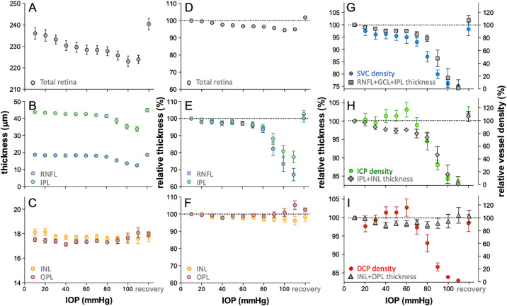
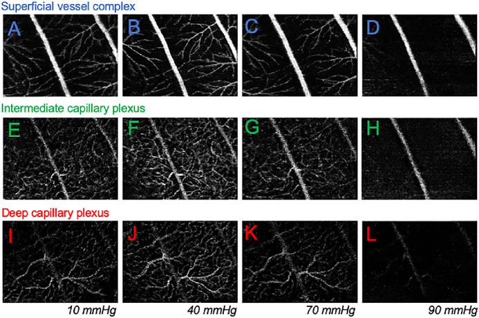
The three vascular layers responded differently to IOP elevation. For IOPs between 40 and 60 mm Hg, DCP and ICP capillaries were significantly more resistant to IOP elevation than those in the SVC. When IOP was elevated above 70 mm Hg, all layers showed reduced vessel density. IOP induced change in SVC vessel density closely followed reductions in thickness of the inner retinal layers (nerve fiber, ganglion cell, and inner plexiform layer). This close relationship between reductions in tissue thickness and vessel density was less apparent for the ICP and DCP.

CONCLUSIONS

These data show that the intermediate and deep vascular plexus in the rat retina have a greater capacity for autoregulation against mild IOP elevation but are more affected at high IOP.

摘要

目的

本研究旨在验证以下假设,即浅层、中层和深层视网膜血管丛对眼内压(IOP)升高的反应不同。

方法

对麻醉的成年 Long Evans 大鼠(n = 14)进行光学相干断层扫描血管造影(OCTA;Spectralis)成像,在基线时(IOP 10mmHg)和在眼压升高(10 至 110mmHg,每步 10mmHg,每步持续 3 分钟)的随访模式下检查血管。拍摄 20°×10°的视野。在二维投影图像中确定浅层血管丛(SVC)、中间毛细血管丛(ICP)和深层毛细血管丛(DCP)内的血管密度(%)。采用 2 路重复测量方差分析(层与 IOP)比较各层之间的差异,然后将结果归一化为基线(相对于 10mmHg 的%相对值)。

结果

这三个血管层对 IOP 升高的反应不同。在 40 至 60mmHg 的 IOP 范围内,DCP 和 ICP 毛细血管对 IOP 升高的抵抗力明显大于 SVC 中的毛细血管。当 IOP 升高到 70mmHg 以上时,所有层的血管密度均降低。SVC 血管密度的 IOP 诱导变化与内视网膜层(神经纤维、节细胞和内丛状层)厚度的减少密切相关。在 ICP 和 DCP 中,这种组织厚度和血管密度减少之间的密切关系不太明显。

结论

这些数据表明,大鼠视网膜的中层和深层血管丛在轻度 IOP 升高时具有更大的自动调节能力,但在高 IOP 时受到的影响更大。

https://cdn.ncbi.nlm.nih.gov/pmc/blobs/f943/7325622/c802f9394147/iovs-61-2-2-f005.jpg
https://cdn.ncbi.nlm.nih.gov/pmc/blobs/f943/7325622/b15cfb20a0bb/iovs-61-2-2-f001.jpg
https://cdn.ncbi.nlm.nih.gov/pmc/blobs/f943/7325622/417e5be0a9a0/iovs-61-2-2-f002.jpg
https://cdn.ncbi.nlm.nih.gov/pmc/blobs/f943/7325622/9687a5f4759e/iovs-61-2-2-f003.jpg
https://cdn.ncbi.nlm.nih.gov/pmc/blobs/f943/7325622/d9aba3886550/iovs-61-2-2-f004.jpg
https://cdn.ncbi.nlm.nih.gov/pmc/blobs/f943/7325622/c802f9394147/iovs-61-2-2-f005.jpg

相似文献

1
Response of the Trilaminar Retinal Vessel Network to Intraocular Pressure Elevation in Rat Eyes.大鼠眼内压升高对三层视网膜血管网络的反应。
Invest Ophthalmol Vis Sci. 2020 Feb 7;61(2):2. doi: 10.1167/iovs.61.2.2.
2
Detection of Reduced Retinal Vessel Density in Eyes with Geographic Atrophy Secondary to Age-Related Macular Degeneration Using Projection-Resolved Optical Coherence Tomography Angiography.采用投影分辨率光学相干断层血管造影术检测与年龄相关性黄斑变性相关的地图状萎缩性眼病的视网膜血管密度降低。
Am J Ophthalmol. 2020 Jan;209:206-212. doi: 10.1016/j.ajo.2019.09.004. Epub 2019 Sep 14.
3
Projection-Resolved Optical Coherence Tomography Angiography of Macular Retinal Circulation in Glaucoma.青光眼黄斑视网膜循环的投影分辨光学相干断层扫描血管造影
Ophthalmology. 2017 Nov;124(11):1589-1599. doi: 10.1016/j.ophtha.2017.06.002. Epub 2017 Jul 1.
4
Deformation of the rodent optic nerve head and peripapillary structures during acute intraocular pressure elevation.在急性眼压升高期间,啮齿动物视神经头部和视盘周围结构的变形。
Invest Ophthalmol Vis Sci. 2011 Aug 22;52(9):6651-61. doi: 10.1167/iovs.11-7578.
5
Relationship between retinal capillary vessel density of OCT angiography and intraocular pressure in pig.猪眼 OCT 血管造影视网膜毛细血管密度与眼压的关系。
Sci Rep. 2021 Apr 20;11(1):8555. doi: 10.1038/s41598-021-87689-8.
6
Analysis of Microvasculature in Nonhuman Primate Macula With Acute Elevated Intraocular Pressure Using Optical Coherence Tomography Angiography.利用光相干断层扫描血管造影术分析急性高眼压非人灵长类动物黄斑区的微血管。
Invest Ophthalmol Vis Sci. 2021 Dec 1;62(15):18. doi: 10.1167/iovs.62.15.18.
7
The Effect of Acutely Elevated Intraocular Pressure on the Functional and Blood Flow Responses of the Rat Retina to Flicker Stimulation.急性眼压升高对大鼠视网膜对闪烁刺激的功能和血流反应的影响。
Invest Ophthalmol Vis Sci. 2017 Oct 1;58(12):5532-5540. doi: 10.1167/iovs.17-22412.
8
Plexus-Specific Retinal Capillary Blood Flow Analysis Using Erythrocyte Mediated Angiography and Optical Coherence Tomography Angiography.应用红细胞介导血管造影和光相干断层扫描血管造影术分析脉络膜特异性视网膜毛细血管血流。
Invest Ophthalmol Vis Sci. 2024 Sep 3;65(11):33. doi: 10.1167/iovs.65.11.33.
9
Glial Cell Contribution to Basal Vessel Diameter and Pressure-Initiated Vascular Responses in Rat Retina.神经胶质细胞对大鼠视网膜基底血管直径及压力引发的血管反应的作用
Invest Ophthalmol Vis Sci. 2017 Jan 1;58(1):1-8. doi: 10.1167/iovs.16-20804.
10
Optical Coherence Tomography Angiography Vessel Density Changes after Acute Intraocular Pressure Elevation.急性眼压升高后光学相干断层扫描血管造影血管密度的变化。
Sci Rep. 2018 Apr 16;8(1):6024. doi: 10.1038/s41598-018-24520-x.

引用本文的文献

1
Rat retinal function attenuation with IOP elevation is impacted by both blood pressure and intracranial pressure.随着眼压升高,大鼠视网膜功能衰减受到血压和颅内压两者的影响。
Front Physiol. 2025 Jun 18;16:1566032. doi: 10.3389/fphys.2025.1566032. eCollection 2025.
2
The Association between Vascular Abnormalities and Glaucoma-What Comes First?血管异常与青光眼之间的关联——哪个为先?
Int J Mol Sci. 2023 Aug 25;24(17):13211. doi: 10.3390/ijms241713211.
3
Retinal Assessment Using In Vivo Electroretinography and Optical Coherence Tomography in Rodent Models of Diabetes.

本文引用的文献

1
Optical Coherence Tomography Angiography in Mice: Quantitative Analysis After Experimental Models of Retinal Damage.光学相干断层扫描血管造影术在小鼠中的应用:视网膜损伤实验模型后的定量分析。
Invest Ophthalmol Vis Sci. 2019 Apr 1;60(5):1556-1565. doi: 10.1167/iovs.18-26441.
2
Optical Coherence Tomography Angiography Vessel Density Changes after Acute Intraocular Pressure Elevation.急性眼压升高后光学相干断层扫描血管造影血管密度的变化。
Sci Rep. 2018 Apr 16;8(1):6024. doi: 10.1038/s41598-018-24520-x.
3
The effects of graded intraocular pressure challenge on the optic nerve head.
在糖尿病啮齿动物模型中使用活体视网膜电图和光学相干断层扫描进行视网膜评估。
Methods Mol Biol. 2023;2678:37-48. doi: 10.1007/978-1-0716-3255-0_4.
4
Intraocular Pressure-Induced Endothelial Dysfunction of Retinal Blood Vessels Is Persistent, but Does Not Trigger Retinal Ganglion Cell Loss.眼压诱导的视网膜血管内皮功能障碍持续存在,但不会引发视网膜神经节细胞丢失。
Antioxidants (Basel). 2022 Sep 21;11(10):1864. doi: 10.3390/antiox11101864.
5
Analysis of Microvasculature in Nonhuman Primate Macula With Acute Elevated Intraocular Pressure Using Optical Coherence Tomography Angiography.利用光相干断层扫描血管造影术分析急性高眼压非人灵长类动物黄斑区的微血管。
Invest Ophthalmol Vis Sci. 2021 Dec 1;62(15):18. doi: 10.1167/iovs.62.15.18.
6
A multi-scale/multi-physics model for the theoretical study of the vascular configuration of retinal capillary plexuses based on OCTA data.一种基于 OCTA 数据的视网膜毛细血管丛血管结构的多尺度/多物理模型的理论研究。
Math Med Biol. 2022 Feb 22;39(1):77-104. doi: 10.1093/imammb/dqab018.
7
Changes of macular vessel density and thickness in gas and silicone oil tamponades after vitrectomy for macula-on rhegmatogenous retinal detachment.孔源性视网膜脱离行玻璃体切除术后黄斑区气体和硅油填充后黄斑区血管密度和厚度的变化。
BMC Ophthalmol. 2021 Nov 15;21(1):392. doi: 10.1186/s12886-021-02160-6.
8
Retrobulbar blood flow in rat eyes during acute elevation of intraocular pressure.大鼠眼内压急性升高时眼后的血流。
Exp Eye Res. 2021 Jun;207:108606. doi: 10.1016/j.exer.2021.108606. Epub 2021 Apr 28.
9
Relationship between retinal capillary vessel density of OCT angiography and intraocular pressure in pig.猪眼 OCT 血管造影视网膜毛细血管密度与眼压的关系。
Sci Rep. 2021 Apr 20;11(1):8555. doi: 10.1038/s41598-021-87689-8.
10
Macular vascular changes in pregnant women with gestational diabetes mellitus by optical coherence tomography angiography.光学相干断层扫描血管造影术观察妊娠期糖尿病孕妇的黄斑血管变化
BMC Ophthalmol. 2021 Apr 9;21(1):170. doi: 10.1186/s12886-021-01927-1.
眼压递增刺激对视神经乳头的影响。
Exp Eye Res. 2018 Apr;169:79-90. doi: 10.1016/j.exer.2018.01.025. Epub 2018 Feb 1.
4
The Effect of Acutely Elevated Intraocular Pressure on the Functional and Blood Flow Responses of the Rat Retina to Flicker Stimulation.急性眼压升高对大鼠视网膜对闪烁刺激的功能和血流反应的影响。
Invest Ophthalmol Vis Sci. 2017 Oct 1;58(12):5532-5540. doi: 10.1167/iovs.17-22412.
5
Ocular fundus pulsations within the posterior rat eye: Chorioscleral motion and response to elevated intraocular pressure.大鼠眼后眼部脉动:脉络膜-巩膜运动和对眼内压升高的反应。
Sci Rep. 2017 Aug 18;7(1):8780. doi: 10.1038/s41598-017-09310-1.
6
The effect of age on the response of retinal capillary filling to changes in intraocular pressure measured by optical coherence tomography angiography.年龄对光学相干断层扫描血管造影测量眼压变化时视网膜毛细血管充盈反应的影响。
Microvasc Res. 2018 Jan;115:12-19. doi: 10.1016/j.mvr.2017.08.001. Epub 2017 Aug 3.
7
Optical coherence tomography angiography: A comprehensive review of current methods and clinical applications.光学相干断层扫描血管造影:当前方法与临床应用的全面综述。
Prog Retin Eye Res. 2017 Sep;60:66-100. doi: 10.1016/j.preteyeres.2017.07.002. Epub 2017 Jul 29.
8
Detailed Vascular Anatomy of the Human Retina by Projection-Resolved Optical Coherence Tomography Angiography.基于层析分辨光学相干断层血管造影术的人视网膜详细血管解剖。
Sci Rep. 2017 Feb 10;7:42201. doi: 10.1038/srep42201.
9
Glial Cell Contribution to Basal Vessel Diameter and Pressure-Initiated Vascular Responses in Rat Retina.神经胶质细胞对大鼠视网膜基底血管直径及压力引发的血管反应的作用
Invest Ophthalmol Vis Sci. 2017 Jan 1;58(1):1-8. doi: 10.1167/iovs.16-20804.
10
Optical coherence tomography angiography of stimulus evoked hemodynamic responses in individual retinal layers.个体视网膜各层刺激诱发血流动力学反应的光学相干断层扫描血管造影术。
Biomed Opt Express. 2016 Jul 29;7(8):3151-62. doi: 10.1364/BOE.7.003151. eCollection 2016 Aug 1.