• 文献检索
  • 文档翻译
  • 深度研究
  • 学术资讯
  • Suppr Zotero 插件Zotero 插件
  • 邀请有礼
  • 套餐&价格
  • 历史记录
应用&插件
Suppr Zotero 插件Zotero 插件浏览器插件Mac 客户端Windows 客户端微信小程序
定价
高级版会员购买积分包购买API积分包
服务
文献检索文档翻译深度研究API 文档MCP 服务
关于我们
关于 Suppr公司介绍联系我们用户协议隐私条款
关注我们

Suppr 超能文献

核心技术专利:CN118964589B侵权必究
粤ICP备2023148730 号-1Suppr @ 2026

文献检索

告别复杂PubMed语法,用中文像聊天一样搜索,搜遍4000万医学文献。AI智能推荐,让科研检索更轻松。

立即免费搜索

文件翻译

保留排版,准确专业,支持PDF/Word/PPT等文件格式,支持 12+语言互译。

免费翻译文档

深度研究

AI帮你快速写综述,25分钟生成高质量综述,智能提取关键信息,辅助科研写作。

立即免费体验

基于机器学习的方法在鉴别 WHO 分级 II 级和 III 级少突胶质细胞瘤方面比放射科医生阅读常规 T1 对比增强和液体衰减反转恢复图像的效果更好。

Better efficacy in differentiating WHO grade II from III oligodendrogliomas with machine-learning than radiologist's reading from conventional T1 contrast-enhanced and fluid attenuated inversion recovery images.

机构信息

Department of Radiology & Functional and Molecular Imaging Key Lab of Shaanxi Province, Tangdu Hospital, Air Force Medical University, 569 Xinsi Road, Xi'an, 710038, Shaanxi, People's Republic of China.

Student Brigade, Air Force Medical University, Xi'an, 710032, Shaanxi, China.

出版信息

BMC Neurol. 2020 Feb 7;20(1):48. doi: 10.1186/s12883-020-1613-y.

DOI:10.1186/s12883-020-1613-y
PMID:32033580
原文链接:https://pmc.ncbi.nlm.nih.gov/articles/PMC7007642/
Abstract

BACKGROUND

The medical imaging to differentiate World Health Organization (WHO) grade II (ODG2) from III (ODG3) oligodendrogliomas still remains a challenge. We investigated whether combination of machine leaning with radiomics from conventional T1 contrast-enhanced (T1 CE) and fluid attenuated inversion recovery (FLAIR) magnetic resonance imaging (MRI) offered superior efficacy.

METHODS

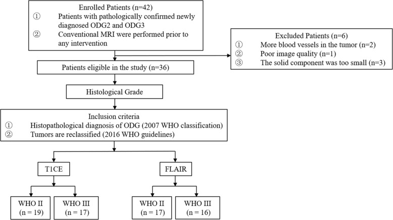
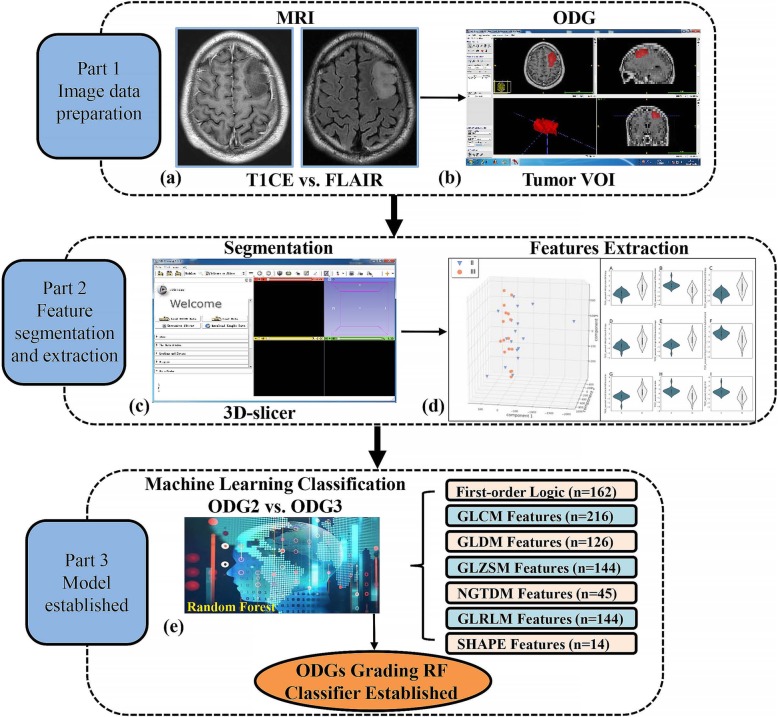
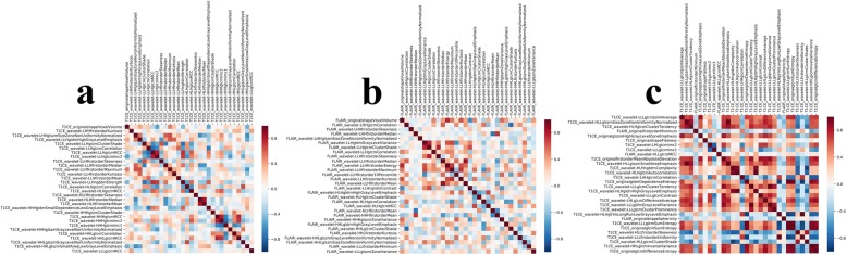
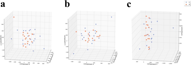
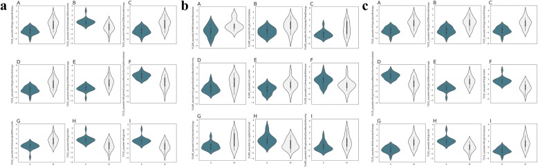
Thirty-six patients with histologically confirmed ODGs underwent T1 CE and 33 of them underwent FLAIR MR examination before any intervention from January 2015 to July 2017 were retrospectively recruited in the current study. The volume of interest (VOI) covering the whole tumor enhancement were manually drawn on the T1 CE and FLAIR slice by slice using ITK-SNAP and a total of 1072 features were extracted from the VOI using 3-D slicer software. Random forest (RF) algorithm was applied to differentiate ODG2 from ODG3 and the efficacy was tested with 5-fold cross validation. The diagnostic efficacy of radiomics-based machine learning and radiologist's assessment were also compared.

RESULTS

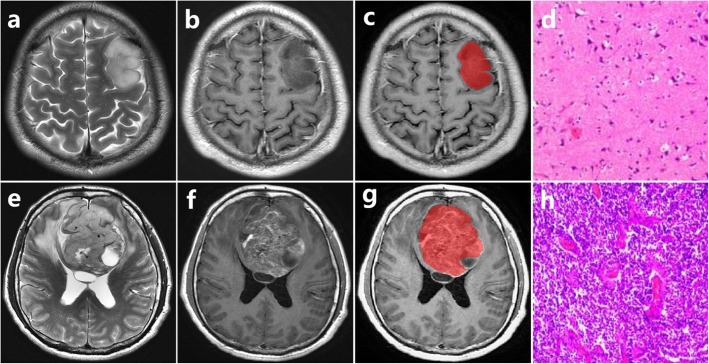
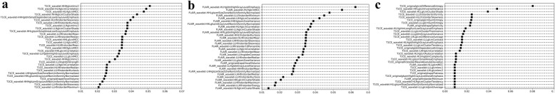
Nineteen ODG2 and 17 ODG3 were included in this study and ODG3 tended to present with prominent necrosis and nodular/ring-like enhancement (P < 0.05). The AUC, ACC, sensitivity, and specificity of radiomics were 0.798, 0.735, 0.672, 0.789 for T1 CE, 0.774, 0.689, 0.700, 0.683 for FLAIR, as well as 0.861, 0.781, 0.778, 0.783 for the combination, respectively. The AUCs of radiologists 1, 2 and 3 were 0.700, 0.687, and 0.714, respectively. The efficacy of machine learning based on radiomics was superior to the radiologists' assessment.

CONCLUSIONS

Machine-learning based on radiomics of T1 CE and FLAIR offered superior efficacy to that of radiologists in differentiating ODG2 from ODG3.

摘要

背景

区分世界卫生组织(WHO)分级 II(ODG2)和 III(ODG3)少突胶质细胞瘤的医学影像学仍然是一个挑战。我们研究了机器学习与常规 T1 对比增强(T1CE)和液体衰减反转恢复(FLAIR)磁共振成像(MRI)的放射组学相结合是否能提供更好的效果。

方法

回顾性招募了 2015 年 1 月至 2017 年 7 月期间接受组织学证实的 ODG 治疗且有 T1CE 检查和 33 例有 FLAIR MRI 检查的 36 例患者。使用 ITK-SNAP 在 T1CE 和 FLAIR 切片上手动绘制覆盖整个肿瘤增强区域的感兴趣区域(VOI),并使用 3D 切片器软件从 VOI 中提取总共 1072 个特征。随机森林(RF)算法用于区分 ODG2 和 ODG3,并使用 5 折交叉验证进行验证。还比较了基于放射组学的机器学习和放射科医生评估的诊断效能。

结果

本研究纳入了 19 例 ODG2 和 17 例 ODG3,ODG3 倾向于表现出明显的坏死和结节状/环状强化(P<0.05)。T1CE 的 AUC、ACC、敏感性和特异性分别为 0.798、0.735、0.672、0.789,FLAIR 为 0.774、0.689、0.700、0.683,联合为 0.861、0.781、0.778、0.783。放射科医生 1、2 和 3 的 AUC 分别为 0.700、0.687 和 0.714。基于放射组学的机器学习的效果优于放射科医生的评估。

结论

T1CE 和 FLAIR 的基于放射组学的机器学习在区分 ODG2 和 ODG3 方面优于放射科医生的评估。

https://cdn.ncbi.nlm.nih.gov/pmc/blobs/ccd3/7007642/a8579a7fc17c/12883_2020_1613_Fig7_HTML.jpg
https://cdn.ncbi.nlm.nih.gov/pmc/blobs/ccd3/7007642/c342b9f6cd2d/12883_2020_1613_Fig1_HTML.jpg
https://cdn.ncbi.nlm.nih.gov/pmc/blobs/ccd3/7007642/354f1f7aabb4/12883_2020_1613_Fig2_HTML.jpg
https://cdn.ncbi.nlm.nih.gov/pmc/blobs/ccd3/7007642/4e4f14907b66/12883_2020_1613_Fig3_HTML.jpg
https://cdn.ncbi.nlm.nih.gov/pmc/blobs/ccd3/7007642/47eb081248f6/12883_2020_1613_Fig4_HTML.jpg
https://cdn.ncbi.nlm.nih.gov/pmc/blobs/ccd3/7007642/5efd4dab959f/12883_2020_1613_Fig5_HTML.jpg
https://cdn.ncbi.nlm.nih.gov/pmc/blobs/ccd3/7007642/d9e02dcf3acb/12883_2020_1613_Fig6_HTML.jpg
https://cdn.ncbi.nlm.nih.gov/pmc/blobs/ccd3/7007642/a8579a7fc17c/12883_2020_1613_Fig7_HTML.jpg

相似文献

1
Better efficacy in differentiating WHO grade II from III oligodendrogliomas with machine-learning than radiologist's reading from conventional T1 contrast-enhanced and fluid attenuated inversion recovery images.基于机器学习的方法在鉴别 WHO 分级 II 级和 III 级少突胶质细胞瘤方面比放射科医生阅读常规 T1 对比增强和液体衰减反转恢复图像的效果更好。
BMC Neurol. 2020 Feb 7;20(1):48. doi: 10.1186/s12883-020-1613-y.
2
Primary central nervous system lymphoma and atypical glioblastoma: Differentiation using radiomics approach.原发性中枢神经系统淋巴瘤和非典型性脑胶质瘤:基于放射组学方法的鉴别诊断。
Eur Radiol. 2018 Sep;28(9):3832-3839. doi: 10.1007/s00330-018-5368-4. Epub 2018 Apr 6.
3
Differentiation of Pseudoprogression from True Progressionin Glioblastoma Patients after Standard Treatment: A Machine Learning Strategy Combinedwith Radiomics Features from T-weighted Contrast-enhanced Imaging.基于 T1 加权对比增强成像的放射组学特征与机器学习策略鉴别胶质母细胞瘤患者标准治疗后假性进展与真性进展。
BMC Med Imaging. 2021 Feb 3;21(1):17. doi: 10.1186/s12880-020-00545-5.
4
Comparison of machine learning classifiers for differentiation of grade 1 from higher gradings in meningioma: A multicenter radiomics study.基于多中心影像组学研究的机器学习分类器在鉴别脑膜瘤 1 级与高级别脑膜瘤中的应用比较。
Magn Reson Imaging. 2019 Nov;63:244-249. doi: 10.1016/j.mri.2019.08.011. Epub 2019 Aug 16.
5
Ability of Radiomics in Differentiation of Anaplastic Oligodendroglioma From Atypical Low-Grade Oligodendroglioma Using Machine-Learning Approach.基于机器学习方法的影像组学在间变性少突胶质细胞瘤与非典型低级别少突胶质细胞瘤鉴别中的应用能力
Front Oncol. 2019 Dec 17;9:1371. doi: 10.3389/fonc.2019.01371. eCollection 2019.
6
An investigation of machine learning methods in delta-radiomics feature analysis.机器学习方法在 delta 放射组学特征分析中的研究。
PLoS One. 2019 Dec 13;14(12):e0226348. doi: 10.1371/journal.pone.0226348. eCollection 2019.
7
Radiomics of Brain MRI: Utility in Prediction of Metastatic Tumor Type.脑 MRI 影像组学:预测转移性肿瘤类型的效用。
Radiology. 2019 Feb;290(2):479-487. doi: 10.1148/radiol.2018180946. Epub 2018 Dec 11.
8
Squamous Cell Carcinoma and Lymphoma of the Oropharynx: Differentiation Using a Radiomics Approach.口咽鳞癌和淋巴瘤:基于放射组学的鉴别方法。
Yonsei Med J. 2020 Oct;61(10):895-900. doi: 10.3349/ymj.2020.61.10.895.
9
Diagnostic Accuracy of a Fluid-attenuated Inversion-Recovery Sequence with Fat Suppression for Assessment of Peripatellar Synovitis: Preliminary Results and Comparison with Contrast-enhanced MR Imaging.带有脂肪抑制的液体衰减反转恢复序列在评估髌周滑膜炎中的诊断准确性:初步结果与对比增强磁共振成像比较。
Radiology. 2017 Jun;283(3):769-778. doi: 10.1148/radiol.2016160155. Epub 2016 Oct 24.
10
Meningiomas: Preoperative predictive histopathological grading based on radiomics of MRI.脑膜瘤:基于 MRI 影像组学的术前预测性组织病理学分级。
Magn Reson Imaging. 2021 Apr;77:36-43. doi: 10.1016/j.mri.2020.11.009. Epub 2020 Nov 18.

引用本文的文献

1
Simplifying Radiomics Workflow for Predicting Grade of Glioma: An Approach for Rapid and Reproducible Radiomics.简化用于预测胶质瘤分级的放射组学工作流程:一种实现快速且可重复的放射组学方法
J Biomed Phys Eng. 2025 Feb 1;15(1):27-36. doi: 10.31661/jbpe.v0i0.2208-1525. eCollection 2025 Feb.
2
Brain tumor grade classification using the ConvNext architecture.使用ConvNext架构进行脑肿瘤分级分类。
Digit Health. 2024 Sep 28;10:20552076241284920. doi: 10.1177/20552076241284920. eCollection 2024 Jan-Dec.
3
Use Test of Automated Machine Learning in Cancer Diagnostics.

本文引用的文献

1
Radiomics Analysis for Glioma Malignancy Evaluation Using Diffusion Kurtosis and Tensor Imaging.基于扩散峰度和张量成像的脑胶质瘤恶性程度评估的放射组学分析。
Int J Radiat Oncol Biol Phys. 2019 Nov 15;105(4):784-791. doi: 10.1016/j.ijrobp.2019.07.011. Epub 2019 Jul 22.
2
Imaging biomarker analysis of advanced multiparametric MRI for glioma grading.高级多参数 MRI 成像生物标志物分析在胶质瘤分级中的应用。
Phys Med. 2019 Apr;60:188-198. doi: 10.1016/j.ejmp.2019.03.014. Epub 2019 Mar 23.
3
Glioma grading using a machine-learning framework based on optimized features obtained from T perfusion MRI and volumes of tumor components.
在癌症诊断中使用自动机器学习测试。
Diagnostics (Basel). 2023 Jul 8;13(14):2315. doi: 10.3390/diagnostics13142315.
4
SWI by 7T MR Imaging for the Microscopic Imaging Diagnosis of Astrocytic and Oligodendroglial Tumors.7T 磁共振扩散峰度成像在星形细胞瘤和少突胶质细胞瘤的微观影像学诊断中的应用。
AJNR Am J Neuroradiol. 2022 Nov;43(11):1575-1581. doi: 10.3174/ajnr.A7666. Epub 2022 Oct 13.
5
Preoperative Diagnosis and Molecular Characterization of Gliomas With Liquid Biopsy and Radiogenomics.基于液体活检和放射基因组学的胶质瘤术前诊断及分子特征分析
Front Neurol. 2022 May 26;13:865171. doi: 10.3389/fneur.2022.865171. eCollection 2022.
6
Fully Automated MR Based Virtual Biopsy of Cerebral Gliomas.基于磁共振成像的脑胶质瘤全自动虚拟活检
Cancers (Basel). 2021 Dec 8;13(24):6186. doi: 10.3390/cancers13246186.
7
Evaluation of the Risk of Recurrence in Patients with Local Advanced Rectal Tumours by Different Radiomic Analysis Approaches.采用不同的放射组学分析方法评估局部晚期直肠肿瘤患者的复发风险
Appl Bionics Biomech. 2021 Nov 28;2021:4520450. doi: 10.1155/2021/4520450. eCollection 2021.
8
Preoperative Radiomics Analysis of 1p/19q Status in WHO Grade II Gliomas.世界卫生组织二级胶质瘤中1p/19q状态的术前影像组学分析
Front Oncol. 2021 Jul 6;11:616740. doi: 10.3389/fonc.2021.616740. eCollection 2021.
9
Accuracy of Machine Learning Algorithms for the Classification of Molecular Features of Gliomas on MRI: A Systematic Literature Review and Meta-Analysis.用于基于MRI对胶质瘤分子特征进行分类的机器学习算法的准确性:一项系统文献综述和荟萃分析
Cancers (Basel). 2021 May 26;13(11):2606. doi: 10.3390/cancers13112606.
10
Diagnostic Accuracy of Machine Learning-Based Radiomics in Grading Gliomas: Systematic Review and Meta-Analysis.基于机器学习的放射组学在胶质瘤分级中的诊断准确性:系统评价和荟萃分析。
Contrast Media Mol Imaging. 2020 Dec 18;2020:2127062. doi: 10.1155/2020/2127062. eCollection 2020.
基于 T 灌注 MRI 优化特征和肿瘤成分体积的机器学习框架进行脑胶质瘤分级。
J Magn Reson Imaging. 2019 Oct;50(4):1295-1306. doi: 10.1002/jmri.26704. Epub 2019 Mar 20.
4
Emerging Applications of Artificial Intelligence in Neuro-Oncology.人工智能在神经肿瘤学中的新兴应用。
Radiology. 2019 Mar;290(3):607-618. doi: 10.1148/radiol.2018181928. Epub 2019 Jan 22.
5
Primary central nervous system lymphoma and atypical glioblastoma: Differentiation using radiomics approach.原发性中枢神经系统淋巴瘤和非典型性脑胶质瘤:基于放射组学方法的鉴别诊断。
Eur Radiol. 2018 Sep;28(9):3832-3839. doi: 10.1007/s00330-018-5368-4. Epub 2018 Apr 6.
6
Radiomics strategy for glioma grading using texture features from multiparametric MRI.基于多参数 MRI 纹理特征的脑胶质瘤分级放射组学策略。
J Magn Reson Imaging. 2018 Dec;48(6):1518-1528. doi: 10.1002/jmri.26010. Epub 2018 Mar 23.
7
MR Imaging of Rectal Cancer: Radiomics Analysis to Assess Treatment Response after Neoadjuvant Therapy.直肠癌磁共振成像:放射组学分析评估新辅助治疗后的治疗反应。
Radiology. 2018 Jun;287(3):833-843. doi: 10.1148/radiol.2018172300. Epub 2018 Mar 7.
8
Artificial Intelligence and Machine Learning in Radiology: Opportunities, Challenges, Pitfalls, and Criteria for Success.人工智能和机器学习在放射学中的应用:机遇、挑战、陷阱和成功标准。
J Am Coll Radiol. 2018 Mar;15(3 Pt B):504-508. doi: 10.1016/j.jacr.2017.12.026. Epub 2018 Feb 4.
9
Distraction in diagnostic radiology: How is search through volumetric medical images affected by interruptions?诊断放射学中的注意力分散:通过容积医学图像进行的搜索是如何受到干扰影响的?
Cogn Res Princ Implic. 2017;2(1):12. doi: 10.1186/s41235-017-0050-y. Epub 2017 Feb 20.
10
ITK-SNAP: An interactive tool for semi-automatic segmentation of multi-modality biomedical images.ITK-SNAP:一种用于多模态生物医学图像半自动分割的交互式工具。
Annu Int Conf IEEE Eng Med Biol Soc. 2016 Aug;2016:3342-3345. doi: 10.1109/EMBC.2016.7591443.