Queen Square Institute of Neurology, University College London, Queen Square 7, London WC1N 3BG, UK.
Lysholm Department of Neuroradiology, The National Hospital for Neurology and Neurosurgery, Queen Square 8-11, London WC1N 3BG, UK.
Contrast Media Mol Imaging. 2020 Dec 18;2020:2127062. doi: 10.1155/2020/2127062. eCollection 2020.
This study aimed to estimate the diagnostic accuracy of machine learning- (ML-) based radiomics in differentiating high-grade gliomas (HGG) from low-grade gliomas (LGG) and to identify potential covariates that could affect the diagnostic accuracy of ML-based radiomic analysis in classifying gliomas.
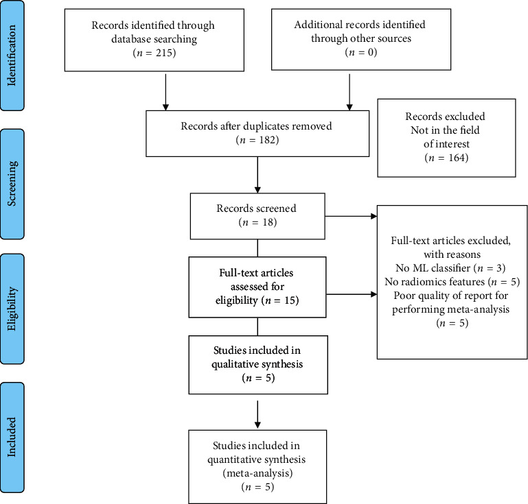
A primary literature search of the PubMed database was conducted to find all related literatures in English between January 1, 2009, and May 1, 2020, with combining synonyms for "machine learning," "glioma," and "radiomics." Five retrospective designed original articles including LGG and HGG subjects were chosen. Pooled sensitivity, specificity, their 95% confidence interval, area under curve (AUC), and hierarchical summary receiver-operating characteristic (HSROC) models were obtained.
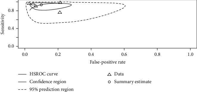
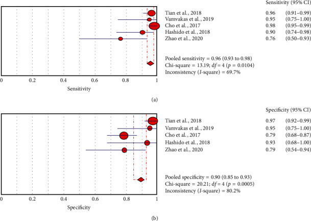
The pooled sensitivity when diagnosing HGG was higher (96% (95% CI: 0.93, 0.98)) than the specificity when diagnosing LGG (90% (95% CI 0.85, 0.93)). Heterogeneity was observed in both sensitivity and specificity. Metaregression confirmed the heterogeneity in sample sizes (=0.05), imaging sequence types (=0.02), and data sources (=0.01), but not for the inclusion of the testing set (=0.19), feature extraction number (=0.36), and selection of feature number (=0.18). The results of subgroup analysis indicate that sample sizes of more than 100 and feature selection numbers less than the total sample size positively affected the diagnostic performance in differentiating HGG from LGG.
This study demonstrates the excellent diagnostic performance of ML-based radiomics in differentiating HGG from LGG.
本研究旨在评估基于机器学习(ML)的放射组学在区分高级别胶质瘤(HGG)和低级别胶质瘤(LGG)方面的诊断准确性,并确定可能影响 ML 基于放射组学分析对胶质瘤进行分类的诊断准确性的潜在协变量。
对 PubMed 数据库进行了初步的文献检索,以查找 2009 年 1 月 1 日至 2020 年 5 月 1 日期间发表的所有相关英文文献,并结合同义词“机器学习”、“胶质瘤”和“放射组学”。选择了 5 篇回顾性设计的原始文章,其中包括 LGG 和 HGG 患者。获得了汇总的敏感性、特异性、95%置信区间、曲线下面积(AUC)和分层综合受试者工作特征(HSROC)模型。
当诊断 HGG 时,汇集的敏感性较高(96%(95%CI:0.93,0.98)),而诊断 LGG 时的特异性较低(90%(95%CI 0.85,0.93))。在敏感性和特异性方面都观察到了异质性。元回归证实了样本量(=0.05)、成像序列类型(=0.02)和数据来源(=0.01)的异质性,但未发现测试集的纳入(=0.19)、特征提取数量(=0.36)和特征选择数量(=0.18)。亚组分析的结果表明,样本量超过 100 个和特征选择数量少于总样本量的特征对区分 HGG 和 LGG 的诊断性能有积极影响。
本研究表明,基于 ML 的放射组学在区分 HGG 和 LGG 方面具有出色的诊断性能。