Turner Institute for Brain and Mental Health, School of Psychological Sciences, Monash University, Melbourne, Victoria, Australia.
Murdoch Children's Research Institute, Melbourne, Victoria, Australia.
Hum Brain Mapp. 2020 May;41(7):1875-1888. doi: 10.1002/hbm.24918. Epub 2020 Feb 7.
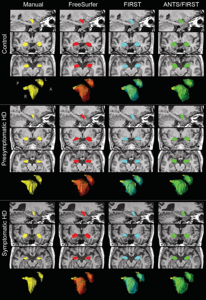
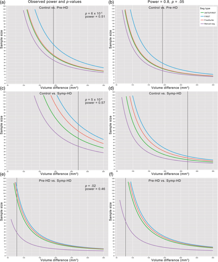
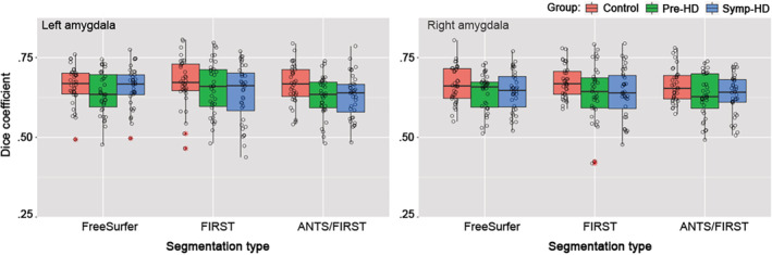
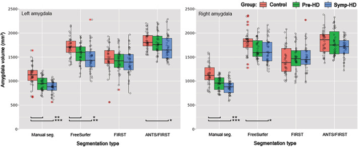
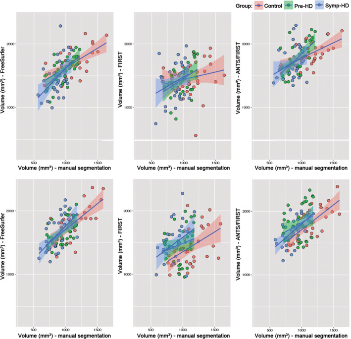
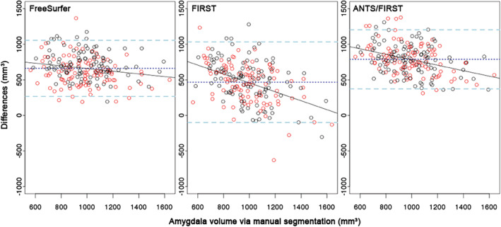
Smaller manually-segmented amygdala volumes have been associated with poorer motor and cognitive function in Huntington's disease (HD). Manual segmentation is the gold standard in terms of accuracy; however, automated methods may be necessary in large samples. Automated segmentation accuracy has not been determined for the amygdala in HD. We aimed to determine which of three automated approaches would most accurately segment amygdalae in HD: FreeSurfer, FIRST, and ANTS nonlinear registration followed by FIRST segmentation. T1-weighted images for the IMAGE-HD cohort including 35 presymptomatic HD (pre-HD), 36 symptomatic HD (symp-HD), and 34 healthy controls were segmented using FreeSurfer and FIRST. For the third approach, images were nonlinearly registered to an MNI template using ANTS, then segmented using FIRST. All automated methods overestimated amygdala volumes compared with manual segmentation. Dice overlap scores, indicating segmentation accuracy, were not significantly different between automated approaches. Manually segmented volumes were most statistically differentiable between groups, followed by those segmented by FreeSurfer, then ANTS/FIRST. FIRST-segmented volumes did not differ between groups. All automated methods produced a bias where volume overestimation was more severe for smaller amygdalae. This bias was subtle for FreeSurfer, but marked for FIRST, and moderate for ANTS/FIRST. Further, FreeSurfer introduced a hemispheric bias not evident with manual segmentation, producing larger right amygdalae by 8%. To assist choice of segmentation approach, we provide sample size estimation graphs based on sample size and other factors. If automated segmentation is employed in samples of the current size, FreeSurfer may effectively distinguish amygdala volume between controls and HD.
较小的手动分割杏仁核体积与亨廷顿病(HD)患者的运动和认知功能较差有关。手动分割在准确性方面是金标准;然而,在大样本中可能需要自动化方法。尚未确定 HD 中杏仁核的自动化分割准确性。我们旨在确定三种自动化方法中的哪一种最能准确分割 HD 中的杏仁核:FreeSurfer、FIRST 和 ANTS 非线性配准后再进行 FIRST 分割。使用 FreeSurfer 和 FIRST 对包括 35 名前驱期 HD(pre-HD)、36 名有症状 HD(symp-HD)和 34 名健康对照的 IMAGE-HD 队列的 T1 加权图像进行分割。对于第三种方法,使用 ANTS 将图像非线性配准到 MNI 模板,然后使用 FIRST 进行分割。与手动分割相比,所有自动化方法都高估了杏仁核体积。指示分割准确性的 Dice 重叠评分在自动化方法之间没有显着差异。手动分割的体积在组间最具统计学差异,其次是 FreeSurfer 分割的体积,然后是 ANTS/FIRST 分割的体积。FIRST 分割的体积在组间没有差异。所有自动化方法都产生了一种偏差,即体积高估在较小的杏仁核中更为严重。FreeSurfer 的这种偏差很细微,但 FIRST 的偏差很明显,而 ANTS/FIRST 的偏差则适中。此外,FreeSurfer 引入了一种手动分割时没有明显显示的半球偏差,导致右侧杏仁核增大 8%。为了帮助选择分割方法,我们根据样本量和其他因素提供了样本量估计图。如果在当前大小的样本中使用自动化分割,FreeSurfer 可能会有效地区分控制组和 HD 之间的杏仁核体积。