Saeed Ramzi Mukred, Dmour Isra, Taha Mutasem O
Department of Pharmaceutical Sciences, Faculty of Pharmacy, University of Jordan, Amman, Jordan.
Faculty of Pharmacy and Medical Sciences, Al-Ahliyya Amman University, Amman, Jordan.
Front Bioeng Biotechnol. 2020 Jan 24;8:4. doi: 10.3389/fbioe.2020.00004. eCollection 2020.
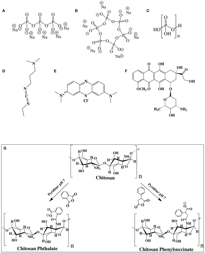
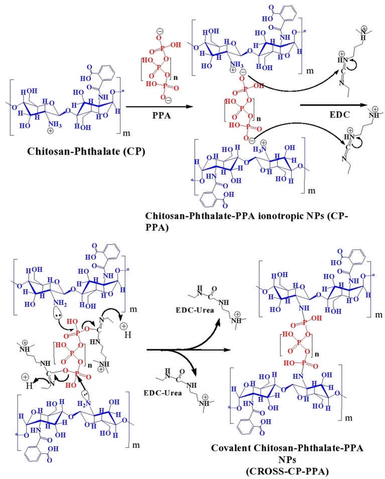
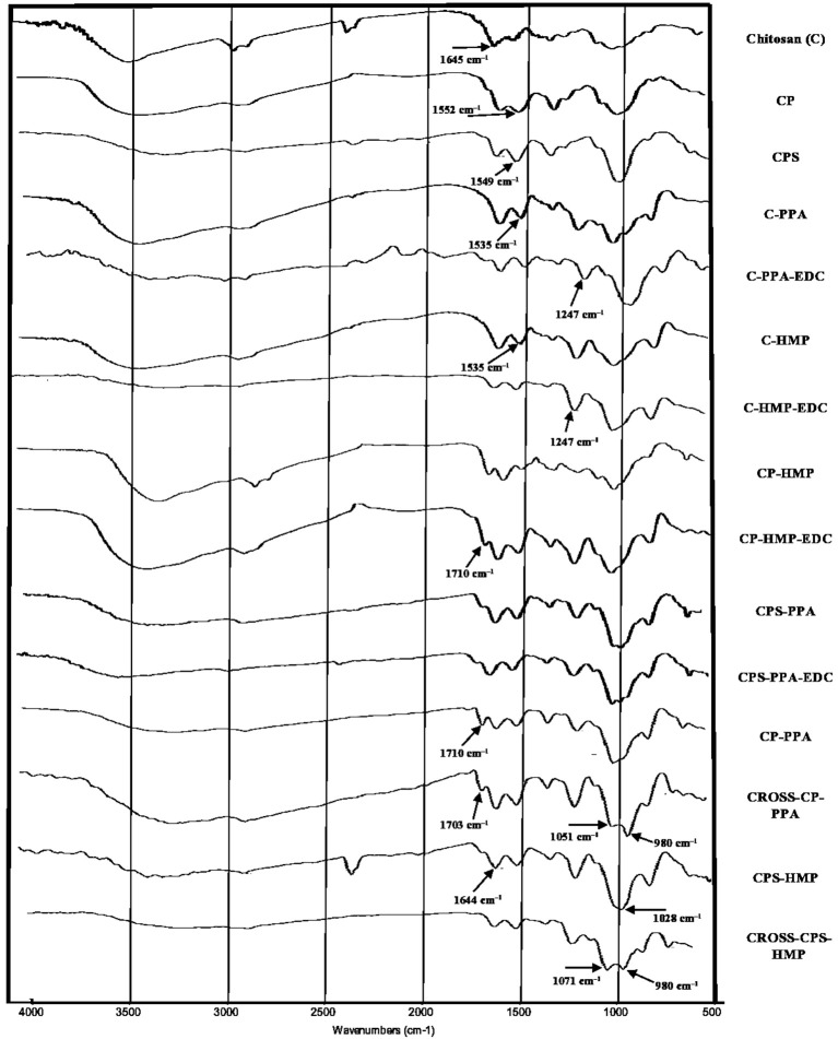
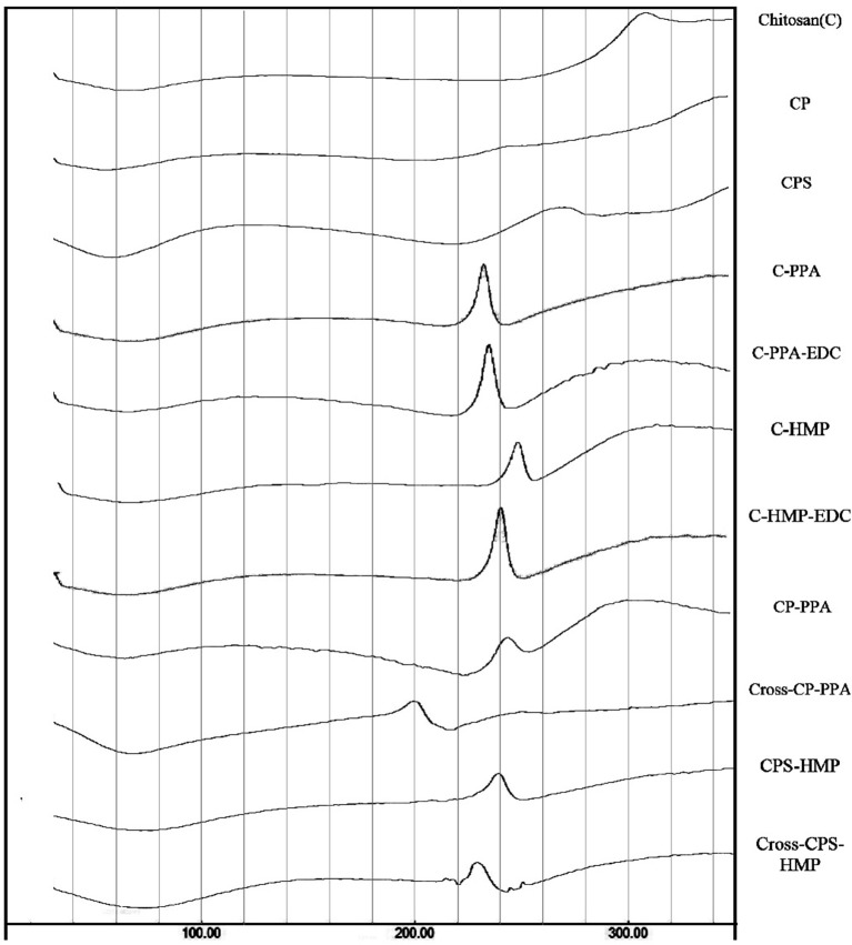
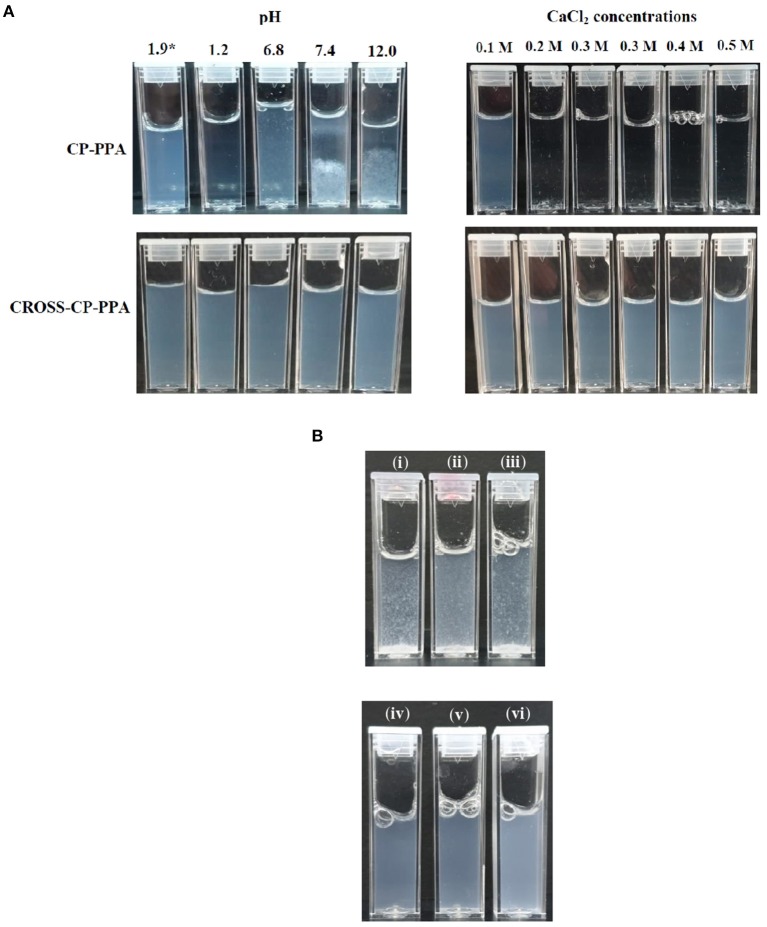
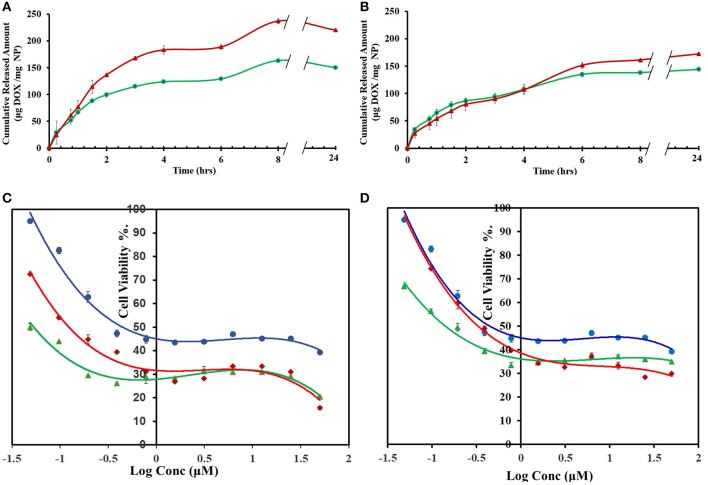
Chitosan nanoparticles (NPs) are widely studied as vehicles for drug, protein, and gene delivery. However, lack of sufficient stability, particularly under physiological conditions, render chitosan NPs of limited pharmaceutical utility. The aim of this study is to produce stable chitosan NPs suitable for drug delivery applications. Chitosan was first grafted to phthalic or phenylsuccinic acids. Subsequently, polyphosphoric acid (PPA), hexametaphosphate (HMP), or tripolyphosphate (TPP) were used to achieve tandem ionotropic/covalently crosslinked chitosan NPs in the presence of 1-ethyl-3-(3-dimethylaminopropyl)-carbodiimide (EDC). Thermal and infrared traits confirmed phosphoramide bonds formation tying chitosan with the polyphosphate crosslinkers within NPs matrices. DLS and TEM size analysis indicated spherical NPs with size range of 120 to 350 nm. The generated NPs exhibited excellent stabilities under harsh pH, CaCl, and 10% FBS conditions. Interestingly, DLS, NPs stability and infrared data suggest HMP to reside within NPs cores, while TPP and PPA to act mainly as NPs surface crosslinkers. Drug loading and release studies using methylene blue (MB) and doxorubicin (DOX) drug models showed covalent PPA- and HMP-based NPs to have superior loading capacities compared to NPs based on unmodified chitosan, generated by ionotropic crosslinking only or covalently crosslinked by TPP. Doxorubicin-loaded NPs were of superior cytotoxic properties against MCF-7 cells compared to free doxorubicin. Specifically, DOX-loaded chitosan-phthalate polyphosphoric acid-crosslinked NPs exhibited 10-folds cytotoxicity enhancement compared to free DOX. The use of PPA and HMP to produce covalently-stabilized chitosan NPs is completely novel.
壳聚糖纳米颗粒(NPs)作为药物、蛋白质和基因递送载体受到广泛研究。然而,缺乏足够的稳定性,尤其是在生理条件下,使得壳聚糖纳米颗粒的药物应用受到限制。本研究的目的是制备适用于药物递送应用的稳定壳聚糖纳米颗粒。壳聚糖首先与邻苯二甲酸或苯琥珀酸接枝。随后,在1-乙基-3-(3-二甲基氨基丙基)-碳二亚胺(EDC)存在下,使用多磷酸(PPA)、六偏磷酸(HMP)或三聚磷酸(TPP)实现串联离子型/共价交联壳聚糖纳米颗粒。热学和红外特性证实了磷酰胺键的形成,将壳聚糖与纳米颗粒基质中的多磷酸盐交联剂连接起来。动态光散射(DLS)和透射电子显微镜(TEM)尺寸分析表明,纳米颗粒呈球形,尺寸范围为120至350nm。所制备的纳米颗粒在苛刻的pH值、氯化钙和10%胎牛血清(FBS)条件下表现出优异的稳定性。有趣的是,DLS、纳米颗粒稳定性和红外数据表明,HMP存在于纳米颗粒核心内,而TPP和PPA主要作为纳米颗粒表面交联剂。使用亚甲蓝(MB)和阿霉素(DOX)药物模型进行的药物负载和释放研究表明,与仅通过离子交联或由TPP共价交联生成的未修饰壳聚糖纳米颗粒相比,基于共价PPA和HMP的纳米颗粒具有更高的负载能力。与游离阿霉素相比,负载阿霉素的纳米颗粒对MCF-7细胞具有更强的细胞毒性。具体而言,与游离DOX相比,负载DOX的壳聚糖-邻苯二甲酸多磷酸交联纳米颗粒的细胞毒性增强了10倍。使用PPA和HMP制备共价稳定的壳聚糖纳米颗粒是全新的方法。