Department of Radiology, Research Institute of Radiological Science, Severance Hospital, Yonsei University College of Medicine, Seoul, South Korea.
Thorac Cancer. 2020 Apr;11(4):993-1004. doi: 10.1111/1759-7714.13352. Epub 2020 Feb 11.
We aimed to assess if quantitative radiomic features can predict programmed death ligand 1 (PD-L1) expression in advanced stage lung adenocarcinoma.
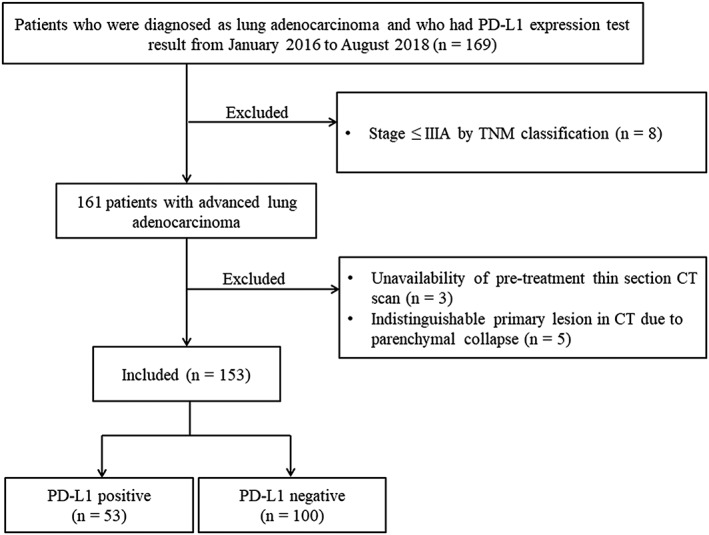
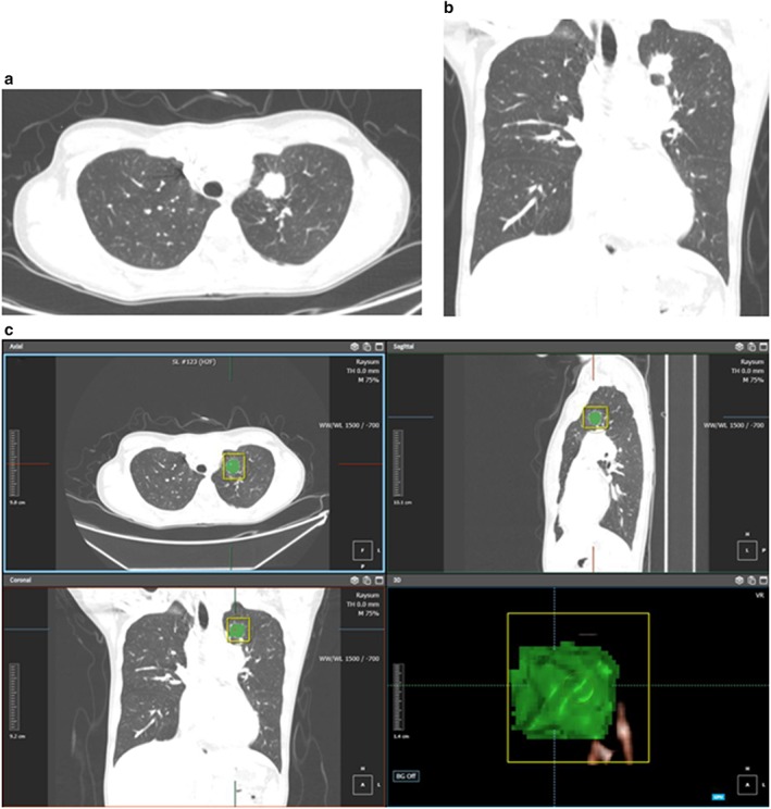
This retrospective study included 153 patients who had advanced stage (>IIIA by TNM classification) lung adenocarcinoma with pretreatment thin section computed tomography (CT) images and PD-L1 expression test results in their pathology reports. Clinicopathological data were collected from electronic medical records. Visual analysis and radiomic feature extraction of the tumor from pretreatment CT were performed. We constructed two models for multivariate logistic regression analysis (one based on clinical variables, and the other based on a combination of clinical variables and radiomic features), and compared c-statistics of the receiver operating characteristic curves of each model to identify the model with the higher predictability.
Among 153 patients, 53 patients were classified as PD-L1 positive and 100 patients as PD-L1 negative. There was no significant difference in clinical characteristics or imaging findings on visual analysis between the two groups (P > 0.05 for all). Rad-score by radiomic analysis was higher in the PD-L1 positive group than in the PD-L1 negative group with a statistical significance (-0.378 ± 1.537 vs. -1.171 ± 0.822, P = 0.0008). A prediction model that uses clinical variables and CT radiomic features showed higher performance compared to a prediction model that uses clinical variables only (c-statistic = 0.646 vs. 0.550, P = 0.0299).
Quantitative CT radiomic features can predict PD-L1 expression in advanced stage lung adenocarcinoma. A prediction model composed of clinical variables and CT radiomic features may facilitate noninvasive assessment of PD-L1 expression.
Significant findings of the study Quantitative CT radiomic features can help predict PD-L1 expression, whereas none of the qualitative imaging findings is associated with PD-L1 positivity. What this study adds A prediction model composed of clinical variables and CT radiomic features may facilitate noninvasive assessment of PD-L1 expression.
本研究旨在评估定量放射组学特征能否预测晚期肺腺癌中程序性死亡配体 1(PD-L1)的表达。
这是一项回顾性研究,纳入了 153 名经病理报告证实为晚期(TNM 分期>IIIA 期)肺腺癌患者,这些患者在治疗前均接受了薄层 CT 扫描,并且其病理报告中提供了 PD-L1 表达检测结果。临床病理数据来自电子病历。我们对治疗前 CT 图像中的肿瘤进行了视觉分析和放射组学特征提取。我们构建了两个多变量逻辑回归分析模型(一个基于临床变量,另一个基于临床变量和放射组学特征的组合),并比较了每个模型的受试者工作特征曲线的 C 统计量,以确定具有更高预测能力的模型。
在 153 名患者中,53 名患者被归类为 PD-L1 阳性,100 名患者为 PD-L1 阴性。两组患者的临床特征或视觉分析影像学表现无显著差异(所有 P 值均>0.05)。放射组学分析的 Rad-score 在 PD-L1 阳性组中高于 PD-L1 阴性组,差异具有统计学意义(-0.378±1.537 比-1.171±0.822,P=0.0008)。与仅使用临床变量的预测模型相比,使用临床变量和 CT 放射组学特征的预测模型具有更高的性能(C 统计量=0.646 比 0.550,P=0.0299)。
定量 CT 放射组学特征可预测晚期肺腺癌中 PD-L1 的表达。由临床变量和 CT 放射组学特征组成的预测模型可能有助于对 PD-L1 表达进行非侵入性评估。
研究的重要发现
定量 CT 放射组学特征有助于预测 PD-L1 的表达,而定性影像学发现均与 PD-L1 阳性无关。
本研究的新增内容
由临床变量和 CT 放射组学特征组成的预测模型可能有助于对 PD-L1 表达进行非侵入性评估。