Kang Dongjie, Qin Zongyuan, Wang Wen, Zheng Yun, Hu Huiying, Bao Yuanyuan, Bao Haihua
Department of Medical Imaging Center, Affiliated Hospital of Qinghai University, Xining, China.
Medicine (Baltimore). 2020 Feb;99(7):e18957. doi: 10.1097/MD.0000000000018957.
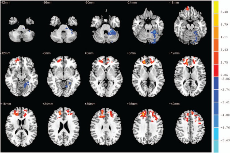
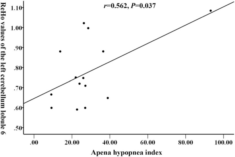
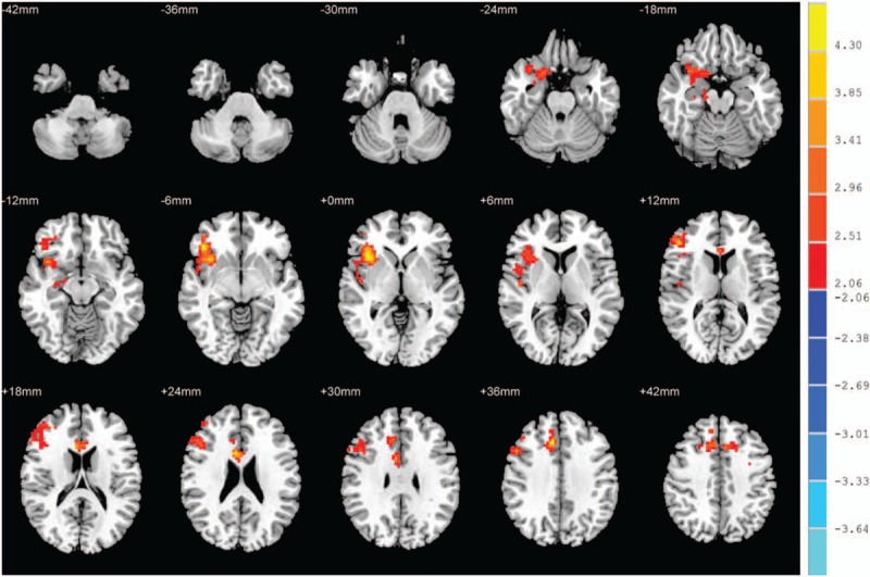
Tibetan is a major ethnic group living on the Qinghai-Tibet Plateau in China. Due to their high-altitude hypoxia environment, sleeping disorder and obstructive sleep apnea hypopnea syndrome (OSAHS) are more prone to occur. In this study, we investigated the brain structural and functional differences between Tibetans OSAHS patients and Tibetans healthy controls using high resolution three-dimensional T1 weighted magnetic resonance imaging (MRI) and resting state functional MRI. The analysis was based on voxel-based morphology, regional homogeneity (ReHo), amplitude of low-frequence fluctuation (ALFF) and functional connection (FC) methods. A total of 14 OSAHS patients and 16 healthy control, all Tibetan male, matched closely in terms of age, education and living altitude, were recruited. The relationship between the ReHo and ALFF values at different brain areas and clinical features, including the apnea hypopnea index (AHI) in the OSAHS group, was analyzed using Pearson correlation. Compared with healthy control, OSAHS patients showed no significant gray matter volume or FC change. OSAHS group showed significantly increased ReHo values in the superior frontal gyrus dorsolateral, the left middle frontal gyrus, and the superior frontal gyrus medial. In contrast, OSAHS group showed decreased ReHo value in the left fusiform gyrus and cerebellum lobule 6. OSAHS group showed significantly increased ALFF values in the right inferior frontal gyrus orbital part, the right median cingulate and paracingulate gyri, the right Inferior frontal gyrus triangular part, the right insula and the left superior frontal gyrus dorsolateral. In the OSAHS group, the AHI showed a positive correlation with the ReHo value at the left cerebellum lobule 6 (r = 0.562, P = .037). Tibetan OSAHS patients had no significant change in brain structure and FC, which may be due to their adaption to the hypoxia environment. ReHo values and ALFF values changes in multiple brain areas in Tibetan OSAHS patients indicated brain functional impairment in multiple brain regions. The left cerebellum lobule 6 gradually compensates brain function as OSAHS progresses.
藏族是中国生活在青藏高原的一个主要民族。由于他们所处的高海拔低氧环境,睡眠障碍和阻塞性睡眠呼吸暂停低通气综合征(OSAHS)更容易发生。在本研究中,我们使用高分辨率三维T1加权磁共振成像(MRI)和静息态功能MRI,研究了藏族OSAHS患者与藏族健康对照者之间的脑结构和功能差异。分析基于基于体素的形态学、局部一致性(ReHo)、低频振幅(ALFF)和功能连接(FC)方法。共招募了14名OSAHS患者和16名健康对照者,均为藏族男性,在年龄、教育程度和居住海拔方面密切匹配。使用Pearson相关性分析了不同脑区的ReHo和ALFF值与临床特征之间的关系,包括OSAHS组的呼吸暂停低通气指数(AHI)。与健康对照相比,OSAHS患者的灰质体积或FC无显著变化。OSAHS组在额上回背外侧、左侧额中回和额上回内侧的ReHo值显著增加。相反,OSAHS组在左侧梭状回和小脑小叶6的ReHo值降低。OSAHS组在右侧额下回眶部、右侧中央扣带回和旁扣带回、右侧额下回三角部、右侧岛叶和左侧额上回背外侧的ALFF值显著增加。在OSAHS组中,AHI与左侧小脑小叶6的ReHo值呈正相关(r = 0.562,P = 0.037)。藏族OSAHS患者的脑结构和FC无显著变化,这可能是由于他们对缺氧环境的适应。藏族OSAHS患者多个脑区的ReHo值和ALFF值变化表明多个脑区存在脑功能损害。随着OSAHS的进展,左侧小脑小叶6逐渐代偿脑功能。