Centre for Heart Lung Innovation, St. Paul's Hospital, Vancouver, BC, Canada.
Department of Anesthesiology, Pharmacology and Therapeutics, University of British Columbia, Vancouver, BC, Canada.
PLoS One. 2020 Feb 14;15(2):e0229278. doi: 10.1371/journal.pone.0229278. eCollection 2020.
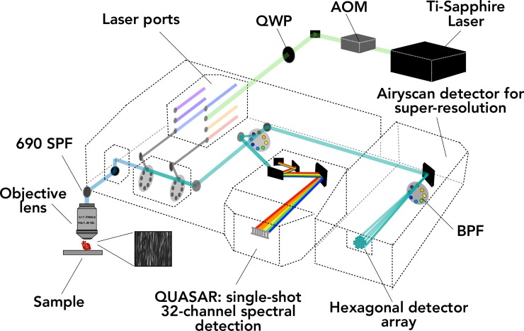
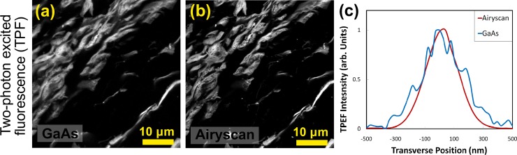
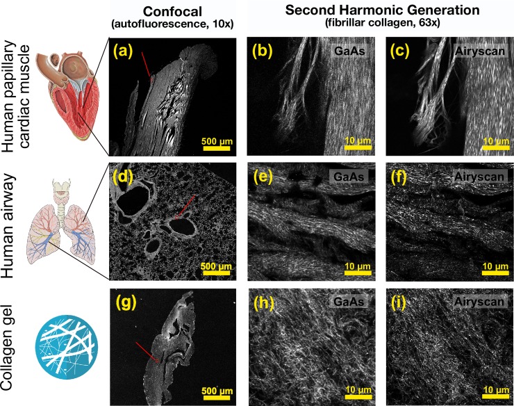
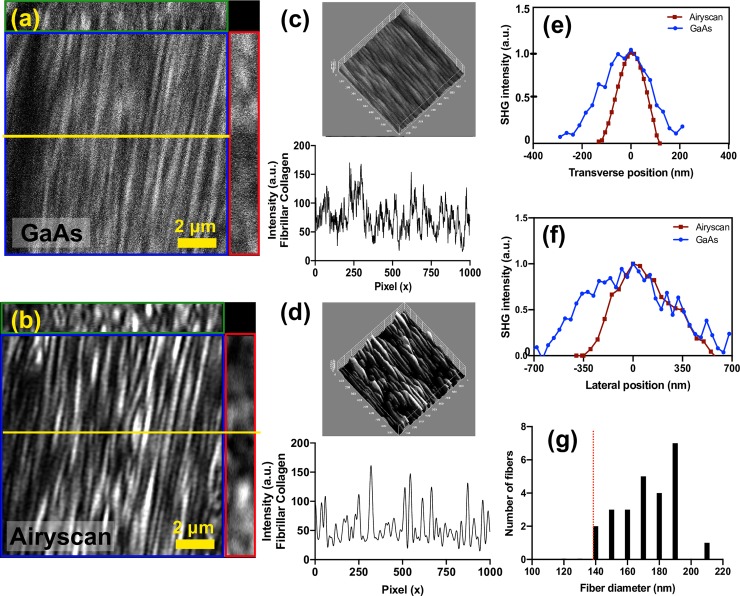
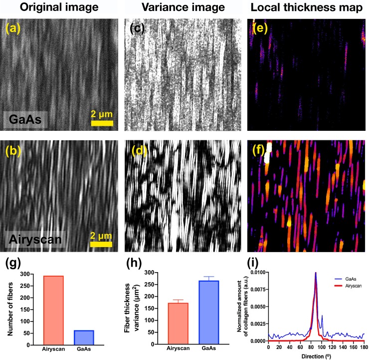
Multiphoton microscopy is a powerful, non-invasive technique to image biological specimens. One current limitation of multiphoton microscopy is resolution as many of the biological molecules and structures investigated by research groups are similar in size or smaller than the diffraction limit. To date, the combination of multiphoton and super-resolution imaging has proved technically challenging for biology focused laboratories to implement. Here we validate that the commercial super-resolution Airyscan detector from ZEISS, which is based on image scanning microscopy, can be integrated under warranty with a pulsed multi-photon laser to enable multiphoton microscopy with super-resolution. We demonstrate its biological application in two different imaging modalities, second harmonic generation (SHG) and two-photon excited fluorescence (TPEF), to measure the fibre thicknesses of collagen and elastin molecules surpassing the diffraction limit by a factor of 1.7±0.3x and 1.4±0.3x respectively, in human heart and lung tissues, and 3-dimensional in vitro models. We show that enhanced resolution and signal-to-noise of SHG using the Airyscan compared to traditional GaAs detectors allows for automated and precise measurement of collagen fibres using texture analysis in biological tissues.
多光子显微镜是一种强大的、非侵入式的技术,用于对生物样本进行成像。目前,多光子显微镜的一个限制是分辨率,因为许多研究小组研究的生物分子和结构的大小相似,或者比衍射极限更小。迄今为止,多光子和超分辨率成像的结合已经被证明对专注于生物学的实验室来说在技术上具有挑战性,难以实施。在这里,我们验证了基于图像扫描显微镜的蔡司商用超分辨率 Airyscan 探测器可以在保修期内与脉冲多光子激光器集成,从而实现具有超分辨率的多光子显微镜。我们在两种不同的成像模式下,即二次谐波产生(SHG)和双光子激发荧光(TPEF),展示了其生物学应用,分别以 1.7±0.3x 和 1.4±0.3x 的因子超越了人类心脏和肺部组织以及体外 3D 模型中胶原和弹性蛋白分子的衍射极限,来测量其纤维厚度。我们表明,与传统的 GaAs 探测器相比,Airyscan 用于 SHG 的增强分辨率和信噪比,可以在生物组织中使用纹理分析对胶原纤维进行自动和精确的测量。