Alrefai M T, Tarola C L, Raagas R, Ridwan K, Shalal M, Lomis N, Paul A, Alrefai M D, Prakash S, Schwertani A, Shum-Tim D
Divisions of Cardiac Surgery and Cardiology, McGill University Health Center, Montreal, QC, Canada.
King Faisal Specialist Hospital and Research Center, Jeddah, Saudi Arabia.
Ann Stem Cell Res. 2019;2(1):29-36. Epub 2019 Nov 7.
Cell-based therapies have demonstrated variable degrees of success in the management of myocardial infarction and heart failure. By inducing a myocardial infarction in a rat model, the effects of secretome from human induced pluripotent stem cells (HiPSCs) and human mesenchymal stem cells (hMSCs) on cardiac function and remodeling were investigated.
HiPSCs and hMSCs were cultured and after 12 cycles, secretome was collected. The quantification of stem cell growth factors was measured using the ELISA test. Thirty female Lewis rats underwent surgical ligation of the left coronary artery. The rats were then randomized (n=10/group) to receive one of three treatments injected into the peri-infarct area; normal saline, HiPSC and hMSC. Left ventricular ejection fraction (LVEF), fractional shortening (FS), histology and serum proteomics were evaluated in a blinded fashion both pre-operatively and at 2, 4 and 6 weeks.
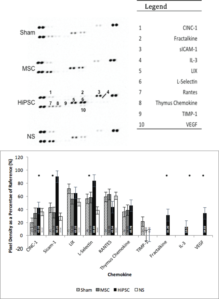
ELISA studies revealed, Platelet-derived growth factor (PDGF) concentration of 3.35± 0.031 ng/ml (0.68± 0.027ng/ml) for MSC-CM group, 3.44± 0.042 ng/ml (0.78± 0.03 ng/ml) for the HiPSC-CM group, 3.2± 0.107 ng/ml (0.64±0.013 ng/ml) for the MSC-pre-group, 3.1± 0.075 ng/ml (0.71± 0.013 ng/ml) for the HiPSC-pre group and 3.3± 0.047 ng/ml (0.71± 0.014ng/ml) for the HiPSC-pre-r group at 60 min in comparison to at (0 min).Compared to non-treated (NT), HiPSC and hMSC, treated rats demonstrated significant improvement in LVEF and FS, and significant reduction in scar size (p<0.05) at 4 and 6 weeks. Proteomic analysis detected the presence of Vascular endothelial growth factor (VEGF) in the serum of rats receiving HiPSC, which was absent in the NT and hMSC groups.
The current study demonstrated a significant improvement of cardiac function and remodeling in response to secretome from HiPSCs and hMSCs. These findings suggest that secretome from HiPSCs may have potential therapy for acute myocardial infarction (MI) without the need of stem cell harvesting and implantation.
基于细胞的疗法在心肌梗死和心力衰竭的治疗中已显示出不同程度的成功。通过在大鼠模型中诱导心肌梗死,研究了人诱导多能干细胞(HiPSC)和人间充质干细胞(hMSC)的分泌组对心脏功能和重塑的影响。
培养HiPSC和hMSC,12个周期后收集分泌组。使用酶联免疫吸附测定(ELISA)试验测量干细胞生长因子的定量。30只雌性Lewis大鼠接受左冠状动脉结扎手术。然后将大鼠随机分组(每组n = 10),在梗死周边区域注射三种治疗方法之一;生理盐水、HiPSC和hMSC。在术前以及术后2、4和6周,以盲法评估左心室射血分数(LVEF)、缩短分数(FS)、组织学和血清蛋白质组学。
ELISA研究显示,与0分钟时相比,MSC-CM组在60分钟时血小板衍生生长因子(PDGF)浓度为3.35±0.031 ng/ml(0.68±0.027 ng/ml),HiPSC-CM组为3.44±0.042 ng/ml(0.78±0.03 ng/ml),MSC-pre组为3.2±0.107 ng/ml(0.64±0.013 ng/ml),HiPSC-pre组为3.1±0.075 ng/ml(0.71±0.013 ng/ml),HiPSC-pre-r组为3.3±0.047 ng/ml(0.71±0.014 ng/ml)。与未治疗(NT)组相比,接受HiPSC和hMSC治疗的大鼠在4周和6周时LVEF和FS有显著改善,瘢痕大小显著减小(p<0.05)。蛋白质组学分析在接受HiPSC治疗的大鼠血清中检测到血管内皮生长因子(VEGF)的存在,而在NT组和hMSC组中未检测到。
当前研究表明,HiPSC和hMSC的分泌组可显著改善心脏功能和重塑。这些发现表明,HiPSC的分泌组可能对急性心肌梗死(MI)具有潜在治疗作用,而无需进行干细胞采集和植入。