• 文献检索
  • 文档翻译
  • 深度研究
  • 学术资讯
  • Suppr Zotero 插件Zotero 插件
  • 邀请有礼
  • 套餐&价格
  • 历史记录
应用&插件
Suppr Zotero 插件Zotero 插件浏览器插件Mac 客户端Windows 客户端微信小程序
定价
高级版会员购买积分包购买API积分包
服务
文献检索文档翻译深度研究API 文档MCP 服务
关于我们
关于 Suppr公司介绍联系我们用户协议隐私条款
关注我们

Suppr 超能文献

核心技术专利:CN118964589B侵权必究
粤ICP备2023148730 号-1Suppr @ 2026

文献检索

告别复杂PubMed语法,用中文像聊天一样搜索,搜遍4000万医学文献。AI智能推荐,让科研检索更轻松。

立即免费搜索

文件翻译

保留排版,准确专业,支持PDF/Word/PPT等文件格式,支持 12+语言互译。

免费翻译文档

深度研究

AI帮你快速写综述,25分钟生成高质量综述,智能提取关键信息,辅助科研写作。

立即免费体验

BSCN:用于视网膜血管分割的双向对称级联网络。

BSCN: bidirectional symmetric cascade network for retinal vessel segmentation.

机构信息

College of Information Science and Engineering,Shandong University of Science and Technology, Shandong, Qingdao 266590, China.

Shandong Province Key Laboratory of Wisdom Mining Information Technology, Shandong University of Science and Technology, Shandong, Qingdao 266590, China.

出版信息

BMC Med Imaging. 2020 Feb 18;20(1):20. doi: 10.1186/s12880-020-0412-7.

DOI:10.1186/s12880-020-0412-7
PMID:32070306
原文链接:https://pmc.ncbi.nlm.nih.gov/articles/PMC7029442/
Abstract

BACKGROUND

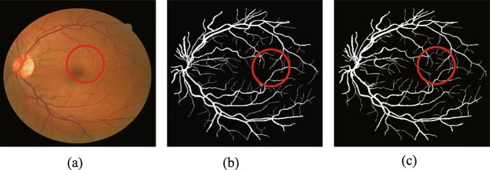
Retinal blood vessel segmentation has an important guiding significance for the analysis and diagnosis of cardiovascular diseases such as hypertension and diabetes. But the traditional manual method of retinal blood vessel segmentation is not only time-consuming and laborious but also cannot guarantee the accuracy and efficiency of diagnosis. Therefore, it is especially significant to create a computer-aided method of automatic and accurate retinal vessel segmentation.

METHODS

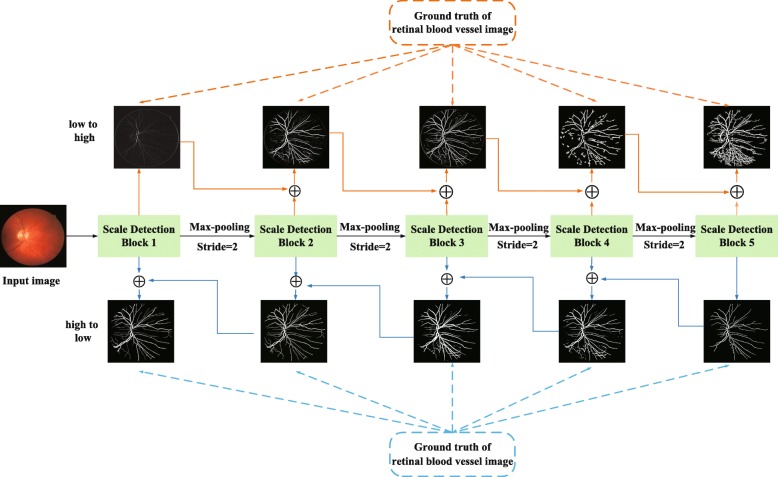
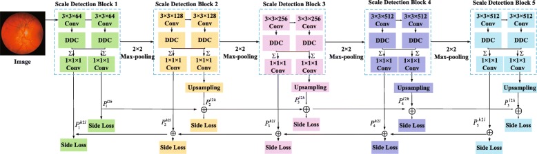
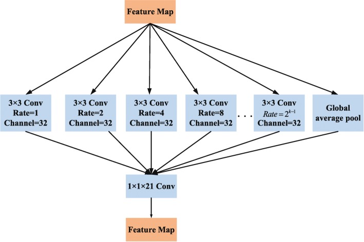
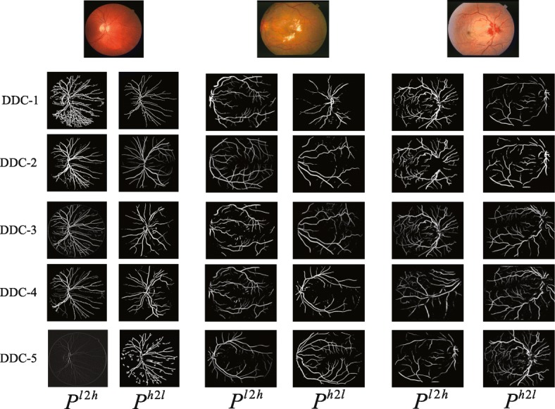
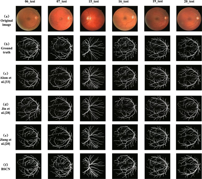
In order to extract the blood vessels' contours of different diameters to realize fine segmentation of retinal vessels, we propose a Bidirectional Symmetric Cascade Network (BSCN) where each layer is supervised by vessel contour labels of specific diameter scale instead of using one general ground truth to train different network layers. In addition, to increase the multi-scale feature representation of retinal blood vessels, we propose the Dense Dilated Convolution Module (DDCM), which extracts retinal vessel features of different diameters by adjusting the dilation rate in the dilated convolution branches and generates two blood vessel contour prediction results by two directions respectively. All dense dilated convolution module outputs are fused to obtain the final vessel segmentation results.

RESULTS

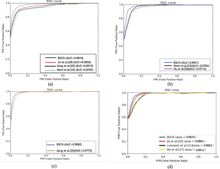
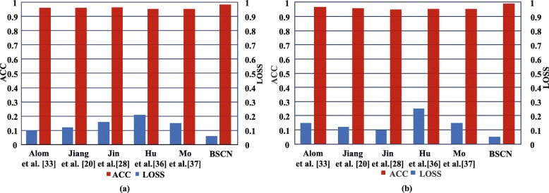
We experimented the three datasets of DRIVE, STARE, HRF and CHASE_DB1, and the proposed method reaches accuracy of 0.9846/0.9872/0.9856/0.9889 and AUC of 0.9874/0.9941/0.9882/0.9874 on DRIVE, STARE, HRF and CHASE_DB1.

CONCLUSIONS

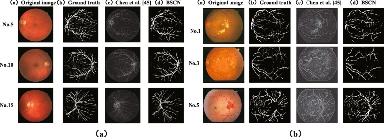
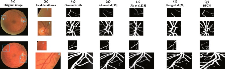
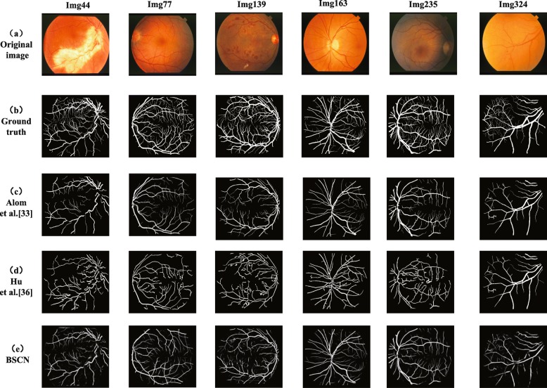
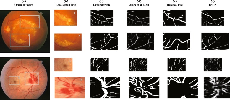
The experimental results show that compared with the state-of-art methods, the proposed method has strong robustness, it not only avoids the adverse interference of the lesion background but also detects the tiny blood vessels at the intersection accurately.

摘要

背景

视网膜血管分割对高血压、糖尿病等心血管疾病的分析和诊断具有重要的指导意义。但是传统的视网膜血管手动分割方法不仅耗时费力,而且不能保证诊断的准确性和效率。因此,创建一种自动、准确的视网膜血管分割的计算机辅助方法尤为重要。

方法

为了提取不同直径的血管轮廓,实现视网膜血管的精细分割,我们提出了一种双向对称级联网络(BSCN),其中每一层都由特定直径尺度的血管轮廓标签进行监督,而不是使用一个通用的真实标签来训练不同的网络层。此外,为了增加视网膜血管的多尺度特征表示,我们提出了密集扩张卷积模块(DDCM),通过调整扩张卷积分支中的扩张率来提取不同直径的视网膜血管特征,并分别从两个方向生成两个血管轮廓预测结果。所有密集扩张卷积模块的输出都融合在一起,得到最终的血管分割结果。

结果

我们在 DRIVE、STARE、HRF 和 CHASE_DB1 三个数据集上进行了实验,所提出的方法在 DRIVE、STARE、HRF 和 CHASE_DB1 上的准确率分别达到了 0.9846/0.9872/0.9856/0.9889,AUC 分别达到了 0.9874/0.9941/0.9882/0.9874。

结论

实验结果表明,与最先进的方法相比,所提出的方法具有较强的鲁棒性,不仅能避免病变背景的不利干扰,还能准确检测到交叉处的微小血管。

https://cdn.ncbi.nlm.nih.gov/pmc/blobs/494e/7029442/0cec108c04c7/12880_2020_412_Fig17_HTML.jpg
https://cdn.ncbi.nlm.nih.gov/pmc/blobs/494e/7029442/b30cbca001fe/12880_2020_412_Fig1_HTML.jpg
https://cdn.ncbi.nlm.nih.gov/pmc/blobs/494e/7029442/536322e05fb6/12880_2020_412_Fig2_HTML.jpg
https://cdn.ncbi.nlm.nih.gov/pmc/blobs/494e/7029442/b8916e0704f5/12880_2020_412_Fig3_HTML.jpg
https://cdn.ncbi.nlm.nih.gov/pmc/blobs/494e/7029442/c554370e9aaa/12880_2020_412_Fig4_HTML.jpg
https://cdn.ncbi.nlm.nih.gov/pmc/blobs/494e/7029442/cba740a1a3b5/12880_2020_412_Fig5_HTML.jpg
https://cdn.ncbi.nlm.nih.gov/pmc/blobs/494e/7029442/8e242eaa9b16/12880_2020_412_Fig6_HTML.jpg
https://cdn.ncbi.nlm.nih.gov/pmc/blobs/494e/7029442/12eca0af8a44/12880_2020_412_Fig7_HTML.jpg
https://cdn.ncbi.nlm.nih.gov/pmc/blobs/494e/7029442/f9ff548fda90/12880_2020_412_Fig8_HTML.jpg
https://cdn.ncbi.nlm.nih.gov/pmc/blobs/494e/7029442/dc669ba2fa4a/12880_2020_412_Fig9_HTML.jpg
https://cdn.ncbi.nlm.nih.gov/pmc/blobs/494e/7029442/f4b536204e74/12880_2020_412_Fig10_HTML.jpg
https://cdn.ncbi.nlm.nih.gov/pmc/blobs/494e/7029442/65d32863d67d/12880_2020_412_Fig11_HTML.jpg
https://cdn.ncbi.nlm.nih.gov/pmc/blobs/494e/7029442/5287aa60934f/12880_2020_412_Fig12_HTML.jpg
https://cdn.ncbi.nlm.nih.gov/pmc/blobs/494e/7029442/0bd644a2e17e/12880_2020_412_Fig13_HTML.jpg
https://cdn.ncbi.nlm.nih.gov/pmc/blobs/494e/7029442/8c6c2c40bc2f/12880_2020_412_Fig14_HTML.jpg
https://cdn.ncbi.nlm.nih.gov/pmc/blobs/494e/7029442/e63f7f36f98b/12880_2020_412_Fig15_HTML.jpg
https://cdn.ncbi.nlm.nih.gov/pmc/blobs/494e/7029442/78409ce2f166/12880_2020_412_Fig16_HTML.jpg
https://cdn.ncbi.nlm.nih.gov/pmc/blobs/494e/7029442/0cec108c04c7/12880_2020_412_Fig17_HTML.jpg

相似文献

1
BSCN: bidirectional symmetric cascade network for retinal vessel segmentation.BSCN:用于视网膜血管分割的双向对称级联网络。
BMC Med Imaging. 2020 Feb 18;20(1):20. doi: 10.1186/s12880-020-0412-7.
2
Retinal blood vessel extraction employing effective image features and combination of supervised and unsupervised machine learning methods.采用有效图像特征和监督与无监督机器学习方法相结合的视网膜血管提取。
Artif Intell Med. 2019 Apr;95:1-15. doi: 10.1016/j.artmed.2019.03.001. Epub 2019 Mar 2.
3
Densely connected U-Net retinal vessel segmentation algorithm based on multi-scale feature convolution extraction.基于多尺度特征卷积提取的密集连接 U-Net 视网膜血管分割算法。
Med Phys. 2021 Jul;48(7):3827-3841. doi: 10.1002/mp.14944. Epub 2021 Jun 16.
4
Retinal blood vessel segmentation using fully convolutional network with transfer learning.基于迁移学习的全卷积网络的视网膜血管分割。
Comput Med Imaging Graph. 2018 Sep;68:1-15. doi: 10.1016/j.compmedimag.2018.04.005. Epub 2018 Apr 26.
5
G2ViT: Graph Neural Network-Guided Vision Transformer Enhanced Network for retinal vessel and coronary angiograph segmentation.G2ViT:基于图神经网络引导的视觉Transformer 增强网络,用于视网膜血管和冠状动脉造影分割。
Neural Netw. 2024 Aug;176:106356. doi: 10.1016/j.neunet.2024.106356. Epub 2024 May 3.
6
MCFSA-Net: A multi-scale channel fusion and spatial activation network for retinal vessel segmentation.MCFSA-Net:一种用于视网膜血管分割的多尺度通道融合与空间激活网络。
J Biophotonics. 2023 Apr;16(4):e202200295. doi: 10.1002/jbio.202200295. Epub 2022 Dec 1.
7
Multiscale U-Net with Spatial Positional Attention for Retinal Vessel Segmentation.多尺度 U-Net 结合空间位置注意力的视网膜血管分割。
J Healthc Eng. 2022 Jan 10;2022:5188362. doi: 10.1155/2022/5188362. eCollection 2022.
8
SegR-Net: A deep learning framework with multi-scale feature fusion for robust retinal vessel segmentation.SegR-Net:一种具有多尺度特征融合的深度学习框架,用于稳健的视网膜血管分割。
Comput Biol Med. 2023 Sep;163:107132. doi: 10.1016/j.compbiomed.2023.107132. Epub 2023 Jun 10.
9
Scale-space approximated convolutional neural networks for retinal vessel segmentation.用于视网膜血管分割的尺度空间逼近卷积神经网络。
Comput Methods Programs Biomed. 2019 Sep;178:237-246. doi: 10.1016/j.cmpb.2019.06.030. Epub 2019 Jun 29.
10
PCAT-UNet: UNet-like network fused convolution and transformer for retinal vessel segmentation.PCAT-UNet:融合卷积和变形注意力的 U 型网络用于视网膜血管分割。
PLoS One. 2022 Jan 24;17(1):e0262689. doi: 10.1371/journal.pone.0262689. eCollection 2022.

引用本文的文献

1
Recent developments on computer aided systems for diagnosis of diabetic retinopathy: a review.糖尿病视网膜病变诊断的计算机辅助系统的最新进展:综述
Multimed Tools Appl. 2023;82(10):14471-14525. doi: 10.1007/s11042-022-13841-9. Epub 2022 Sep 24.
2
Practical utility of liver segmentation methods in clinical surgeries and interventions.肝脏分割方法在临床手术和介入中的实际应用。
BMC Med Imaging. 2022 May 24;22(1):97. doi: 10.1186/s12880-022-00825-2.
3
State-of-the-art retinal vessel segmentation with minimalistic models.基于极简模型的视网膜血管分割技术的最新进展。

本文引用的文献

1
CE-Net: Context Encoder Network for 2D Medical Image Segmentation.CE-Net:用于二维医学图像分割的上下文编码器网络。
IEEE Trans Med Imaging. 2019 Oct;38(10):2281-2292. doi: 10.1109/TMI.2019.2903562. Epub 2019 Mar 7.
2
Retinal blood vessel segmentation using fully convolutional network with transfer learning.基于迁移学习的全卷积网络的视网膜血管分割。
Comput Med Imaging Graph. 2018 Sep;68:1-15. doi: 10.1016/j.compmedimag.2018.04.005. Epub 2018 Apr 26.
3
Universal eye health: are we getting closer?全球眼健康:我们离目标更近了吗?
Sci Rep. 2022 Apr 13;12(1):6174. doi: 10.1038/s41598-022-09675-y.
4
Retinal Vessel Automatic Segmentation Using SegNet.基于 SegNet 的视网膜血管自动分割。
Comput Math Methods Med. 2022 Mar 26;2022:3117455. doi: 10.1155/2022/3117455. eCollection 2022.
5
Diabetic and Hypertensive Retinopathy Screening in Fundus Images Using Artificially Intelligent Shallow Architectures.利用人工智能浅层架构进行眼底图像中的糖尿病性和高血压性视网膜病变筛查。
J Pers Med. 2021 Dec 23;12(1):7. doi: 10.3390/jpm12010007.
Lancet Glob Health. 2017 Sep;5(9):e843-e844. doi: 10.1016/S2214-109X(17)30302-9. Epub 2017 Aug 2.
4
Multi-level deep supervised networks for retinal vessel segmentation.多层深度监督网络用于视网膜血管分割。
Int J Comput Assist Radiol Surg. 2017 Dec;12(12):2181-2193. doi: 10.1007/s11548-017-1619-0. Epub 2017 Jun 2.
5
Development and Validation of a Deep Learning Algorithm for Detection of Diabetic Retinopathy in Retinal Fundus Photographs.深度学习算法在视网膜眼底照片糖尿病视网膜病变检测中的开发与验证。
JAMA. 2016 Dec 13;316(22):2402-2410. doi: 10.1001/jama.2016.17216.
6
Segmenting Retinal Blood Vessels With Deep Neural Networks.基于深度神经网络的视网膜血管分割。
IEEE Trans Med Imaging. 2016 Nov;35(11):2369-2380. doi: 10.1109/TMI.2016.2546227. Epub 2016 Mar 24.
7
Curve-Like Structure Extraction Using Minimal Path Propagation With Backtracking.使用带回溯的最小路径传播进行曲线状结构提取。
IEEE Trans Image Process. 2016 Feb;25(2):988-1003. doi: 10.1109/TIP.2015.2496279. Epub 2015 Nov 2.
8
Vessel extraction from non-fluorescein fundus images using orientation-aware detector.使用方向感知检测器从非荧光眼底图像中提取血管。
Med Image Anal. 2015 Dec;26(1):232-42. doi: 10.1016/j.media.2015.09.002. Epub 2015 Sep 25.
9
A Graph-Theoretical Approach for Tracing Filamentary Structures in Neuronal and Retinal Images.一种用于追踪神经元和视网膜图像中丝状结构的图论方法。
IEEE Trans Med Imaging. 2016 Jan;35(1):257-72. doi: 10.1109/TMI.2015.2465962. Epub 2015 Aug 24.
10
Trainable COSFIRE filters for vessel delineation with application to retinal images.可训练的 COSFIRE 滤波器在视网膜图像中的血管分割应用
Med Image Anal. 2015 Jan;19(1):46-57. doi: 10.1016/j.media.2014.08.002. Epub 2014 Sep 3.