• 文献检索
  • 文档翻译
  • 深度研究
  • 学术资讯
  • Suppr Zotero 插件Zotero 插件
  • 邀请有礼
  • 套餐&价格
  • 历史记录
应用&插件
Suppr Zotero 插件Zotero 插件浏览器插件Mac 客户端Windows 客户端微信小程序
定价
高级版会员购买积分包购买API积分包
服务
文献检索文档翻译深度研究API 文档MCP 服务
关于我们
关于 Suppr公司介绍联系我们用户协议隐私条款
关注我们

Suppr 超能文献

核心技术专利:CN118964589B侵权必究
粤ICP备2023148730 号-1Suppr @ 2026

文献检索

告别复杂PubMed语法,用中文像聊天一样搜索,搜遍4000万医学文献。AI智能推荐,让科研检索更轻松。

立即免费搜索

文件翻译

保留排版,准确专业,支持PDF/Word/PPT等文件格式,支持 12+语言互译。

免费翻译文档

深度研究

AI帮你快速写综述,25分钟生成高质量综述,智能提取关键信息,辅助科研写作。

立即免费体验

基于 SegNet 的视网膜血管自动分割。

Retinal Vessel Automatic Segmentation Using SegNet.

机构信息

School of Medical Technology and Information Engineering, Zhejiang Chinese Medical University, Hangzhou 310053, China.

出版信息

Comput Math Methods Med. 2022 Mar 26;2022:3117455. doi: 10.1155/2022/3117455. eCollection 2022.

DOI:10.1155/2022/3117455
PMID:35378728
原文链接:https://pmc.ncbi.nlm.nih.gov/articles/PMC8976667/
Abstract

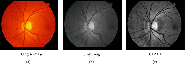
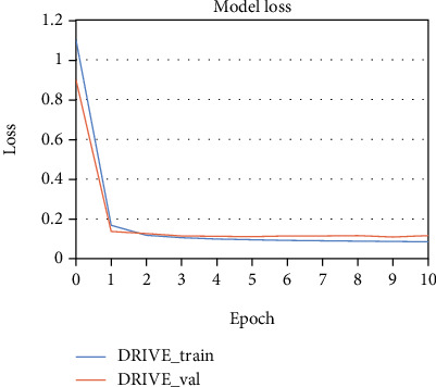
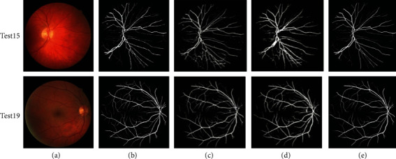
Extracting retinal vessels accurately is very important for diagnosing some diseases such as diabetes retinopathy, hypertension, and cardiovascular. Clinically, experienced ophthalmologists diagnose these diseases through segmenting retinal vessels manually and analysing its structural feature, such as tortuosity and diameter. However, manual segmentation of retinal vessels is a time-consuming and laborious task with strong subjectivity. The automatic segmentation technology of retinal vessels can not only reduce the burden of ophthalmologists but also effectively solve the problem that is a lack of experienced ophthalmologists in remote areas. Therefore, the automatic segmentation technology of retinal vessels is of great significance for clinical auxiliary diagnosis and treatment of ophthalmic diseases. A method using SegNet is proposed in this paper to improve the accuracy of the retinal vessel segmentation. The performance of the retinal vessel segmentation model with SegNet is evaluated on the three public datasets (DRIVE, STARE, and HRF) and achieved accuracy of 0.9518, 0.9683, and 0.9653, sensitivity of 0.7580, 0.7747, and 0.7070, specificity of 0.9804, 0.9910, and 0.9885, score of 0.7992, 0.8369, and 0.7918, MCC of 0.7749, 0.8227, and 0.7643, and AUC of 0.9750, 0.9893, and 0.9740, respectively. The experimental results showed that the method proposed in this research presented better results than many classical methods studied and may be expected to have clinical application prospects.

摘要

准确提取视网膜血管对于诊断糖尿病视网膜病变、高血压和心血管等疾病非常重要。临床上,经验丰富的眼科医生通过手动分割视网膜血管并分析其结构特征(如迂曲度和直径)来诊断这些疾病。然而,视网膜血管的手动分割是一项耗时费力且主观性很强的任务。视网膜血管的自动分割技术不仅可以减轻眼科医生的负担,还可以有效解决偏远地区缺乏经验丰富的眼科医生的问题。因此,视网膜血管的自动分割技术对于眼科疾病的临床辅助诊断和治疗具有重要意义。本文提出了一种使用 SegNet 的方法来提高视网膜血管分割的准确性。在三个公共数据集(DRIVE、STARE 和 HRF)上评估了使用 SegNet 的视网膜血管分割模型的性能,达到了 0.9518、0.9683 和 0.9653 的准确率,0.7580、0.7747 和 0.7070 的敏感度,0.9804、0.9910 和 0.9885 的特异性,0.7992、0.8369 和 0.7918 的得分,0.7749、0.8227 和 0.7643 的 MCC,0.9750、0.9893 和 0.9740 的 AUC。实验结果表明,本研究提出的方法比许多经典方法的研究结果更好,有望具有临床应用前景。

https://cdn.ncbi.nlm.nih.gov/pmc/blobs/ee7c/8976667/072fcaac6dca/CMMM2022-3117455.011.jpg
https://cdn.ncbi.nlm.nih.gov/pmc/blobs/ee7c/8976667/a8a9de15df4e/CMMM2022-3117455.001.jpg
https://cdn.ncbi.nlm.nih.gov/pmc/blobs/ee7c/8976667/6edecd1c70a1/CMMM2022-3117455.002.jpg
https://cdn.ncbi.nlm.nih.gov/pmc/blobs/ee7c/8976667/a8aad801b8c3/CMMM2022-3117455.003.jpg
https://cdn.ncbi.nlm.nih.gov/pmc/blobs/ee7c/8976667/c5b535b80322/CMMM2022-3117455.004.jpg
https://cdn.ncbi.nlm.nih.gov/pmc/blobs/ee7c/8976667/7ec717cab68a/CMMM2022-3117455.005.jpg
https://cdn.ncbi.nlm.nih.gov/pmc/blobs/ee7c/8976667/afe892df3487/CMMM2022-3117455.006.jpg
https://cdn.ncbi.nlm.nih.gov/pmc/blobs/ee7c/8976667/54fe872f2c4e/CMMM2022-3117455.007.jpg
https://cdn.ncbi.nlm.nih.gov/pmc/blobs/ee7c/8976667/a51caadaa553/CMMM2022-3117455.008.jpg
https://cdn.ncbi.nlm.nih.gov/pmc/blobs/ee7c/8976667/1a62e1d46a6a/CMMM2022-3117455.009.jpg
https://cdn.ncbi.nlm.nih.gov/pmc/blobs/ee7c/8976667/899563e1d2ce/CMMM2022-3117455.010.jpg
https://cdn.ncbi.nlm.nih.gov/pmc/blobs/ee7c/8976667/072fcaac6dca/CMMM2022-3117455.011.jpg

相似文献

1
Retinal Vessel Automatic Segmentation Using SegNet.基于 SegNet 的视网膜血管自动分割。
Comput Math Methods Med. 2022 Mar 26;2022:3117455. doi: 10.1155/2022/3117455. eCollection 2022.
2
BSCN: bidirectional symmetric cascade network for retinal vessel segmentation.BSCN:用于视网膜血管分割的双向对称级联网络。
BMC Med Imaging. 2020 Feb 18;20(1):20. doi: 10.1186/s12880-020-0412-7.
3
An improved supervised and attention mechanism-based U-Net algorithm for retinal vessel segmentation.基于改进的监督和注意力机制的 U-Net 算法用于视网膜血管分割。
Comput Biol Med. 2024 Jan;168:107770. doi: 10.1016/j.compbiomed.2023.107770. Epub 2023 Dec 2.
4
Unfolded deep kernel estimation-attention UNet-based retinal image segmentation.基于展开深层核估计-注意力 UNet 的视网膜图像分割。
Sci Rep. 2023 Nov 24;13(1):20712. doi: 10.1038/s41598-023-48039-y.
5
An automated method for accurate vessel segmentation.一种用于精确血管分割的自动化方法。
Phys Med Biol. 2017 May 7;62(9):3757-3778. doi: 10.1088/1361-6560/aa6418. Epub 2017 Apr 6.
6
Segmentation of retinal blood vessels by a novel hybrid technique- Principal Component Analysis (PCA) and Contrast Limited Adaptive Histogram Equalization (CLAHE).基于主成分分析(PCA)和对比度受限自适应直方图均衡化(CLAHE)的新型混合技术对视网膜血管的分割。
Microvasc Res. 2023 Jul;148:104477. doi: 10.1016/j.mvr.2023.104477. Epub 2023 Feb 4.
7
HDC-Net: A hierarchical dilation convolutional network for retinal vessel segmentation.HDC-Net:一种用于视网膜血管分割的分层扩张卷积网络。
PLoS One. 2021 Sep 7;16(9):e0257013. doi: 10.1371/journal.pone.0257013. eCollection 2021.
8
Improving dense conditional random field for retinal vessel segmentation by discriminative feature learning and thin-vessel enhancement.通过判别特征学习和细血管增强改进用于视网膜血管分割的密集条件随机场
Comput Methods Programs Biomed. 2017 Sep;148:13-25. doi: 10.1016/j.cmpb.2017.06.016. Epub 2017 Jun 24.
9
A new robust method for blood vessel segmentation in retinal fundus images based on weighted line detector and hidden Markov model.基于加权线检测器和隐马尔可夫模型的视网膜眼底图像血管分割新方法。
Comput Methods Programs Biomed. 2020 Apr;187:105231. doi: 10.1016/j.cmpb.2019.105231. Epub 2019 Nov 23.
10
MTNet: A combined diagnosis algorithm of vessel segmentation and diabetic retinopathy for retinal images.MTNet:视网膜图像的血管分割和糖尿病性视网膜病变联合诊断算法。
PLoS One. 2022 Nov 23;17(11):e0278126. doi: 10.1371/journal.pone.0278126. eCollection 2022.

引用本文的文献

1
Retracted: Retinal Vessel Automatic Segmentation Using SegNet.已撤回:使用SegNet进行视网膜血管自动分割
Comput Math Methods Med. 2023 Oct 18;2023:9828704. doi: 10.1155/2023/9828704. eCollection 2023.
2
TiM-Net: Transformer in M-Net for Retinal Vessel Segmentation.TiM-Net:用于视网膜血管分割的 M-Net 中的 Transformer。
J Healthc Eng. 2022 Jul 11;2022:9016401. doi: 10.1155/2022/9016401. eCollection 2022.

本文引用的文献

1
Role of pattern electroretinogram in assessment of retinal dysfunction in hypertensive patients.视网膜图形视网膜电图在评估高血压患者视网膜功能障碍中的作用。
Int J Neurosci. 2023 Jul;133(7):791-796. doi: 10.1080/00207454.2021.1984237. Epub 2021 Oct 6.
2
A novel multiple subdivision-based algorithm for quantitative assessment of retinal vascular tortuosity.一种新的基于多重细分的视网膜血管迂曲定量评估算法。
Exp Biol Med (Maywood). 2021 Oct;246(20):2222-2229. doi: 10.1177/15353702211032898. Epub 2021 Jul 25.
3
A Hybrid Unsupervised Approach for Retinal Vessel Segmentation.
一种混合无监督的视网膜血管分割方法。
Biomed Res Int. 2020 Dec 10;2020:8365783. doi: 10.1155/2020/8365783. eCollection 2020.
4
NFN+: A novel network followed network for retinal vessel segmentation.NFN+:一种新型的网络跟随网络用于视网膜血管分割。
Neural Netw. 2020 Jun;126:153-162. doi: 10.1016/j.neunet.2020.02.018. Epub 2020 Mar 4.
5
BSCN: bidirectional symmetric cascade network for retinal vessel segmentation.BSCN:用于视网膜血管分割的双向对称级联网络。
BMC Med Imaging. 2020 Feb 18;20(1):20. doi: 10.1186/s12880-020-0412-7.
6
A new robust method for blood vessel segmentation in retinal fundus images based on weighted line detector and hidden Markov model.基于加权线检测器和隐马尔可夫模型的视网膜眼底图像血管分割新方法。
Comput Methods Programs Biomed. 2020 Apr;187:105231. doi: 10.1016/j.cmpb.2019.105231. Epub 2019 Nov 23.
7
The Interrelationship between Refractive Error, Blood Vessel Anatomy, and Glaucomatous Visual Field Loss.屈光不正、血管解剖结构与青光眼性视野缺损之间的相互关系。
Transl Vis Sci Technol. 2018 Jan 18;7(1):4. doi: 10.1167/tvst.7.1.4. eCollection 2018 Jan.
8
Ametropia, retinal anatomy, and OCT abnormality patterns in glaucoma. 1. Impacts of refractive error and interartery angle.屈光不正、视网膜解剖和青光眼的 OCT 异常模式。1. 屈光不正和动脉间夹角的影响。
J Biomed Opt. 2017 Dec;22(12):1-11. doi: 10.1117/1.JBO.22.12.121713.
9
SegNet: A Deep Convolutional Encoder-Decoder Architecture for Image Segmentation.SegNet:一种用于图像分割的深度卷积编解码器架构。
IEEE Trans Pattern Anal Mach Intell. 2017 Dec;39(12):2481-2495. doi: 10.1109/TPAMI.2016.2644615. Epub 2017 Jan 2.
10
Correction: A Morphological Hessian Based Approach for Retinal Blood Vessels Segmentation and Denoising Using Region Based Otsu Thresholding.更正:一种基于形态学黑塞矩阵的方法,用于使用基于区域的大津阈值法进行视网膜血管分割和去噪。
PLoS One. 2016 Sep 1;11(9):e0162581. doi: 10.1371/journal.pone.0162581. eCollection 2016.