Luginbuehl Leonie H, El-Sharnouby Sherif, Wang Na, Hibberd Julian M
Department of Plant Sciences University of Cambridge Cambridge UK.
Plant Direct. 2020 Feb 11;4(2):e00188. doi: 10.1002/pld3.188. eCollection 2020 Feb.
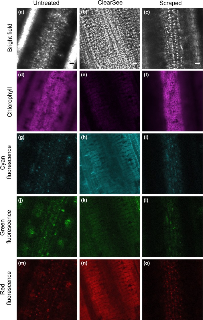
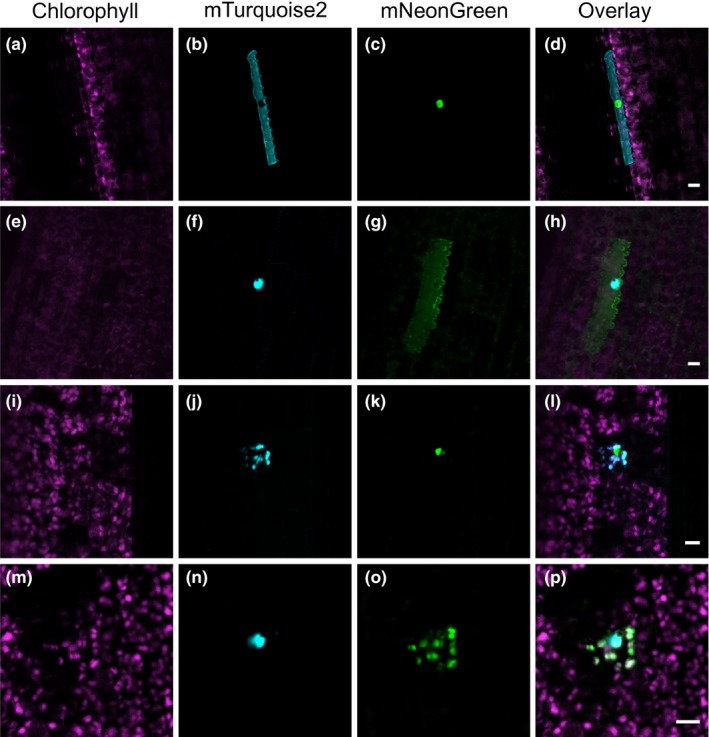
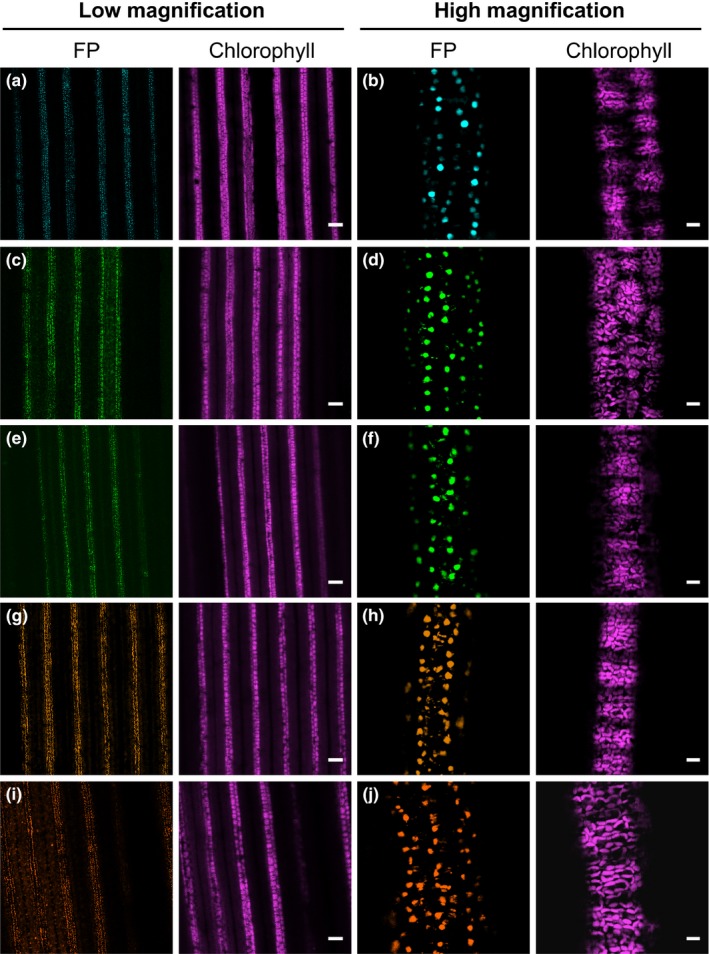
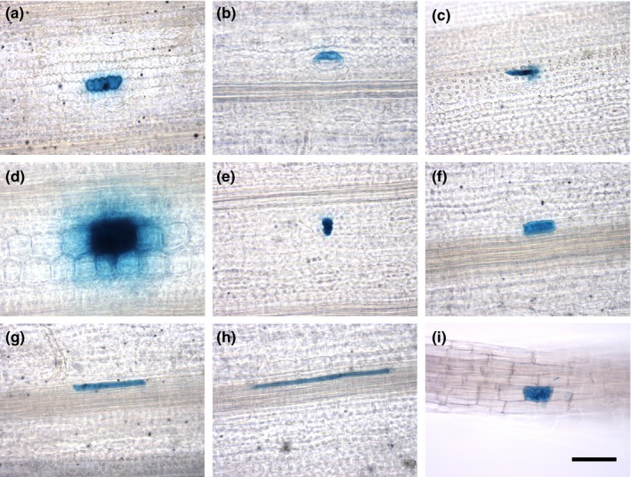
Fluorescent reporters have facilitated non-invasive imaging in multiple plant species and thus allowed the analysis of processes ranging from gene expression and protein localization to cellular patterning. However, in rice, a globally important crop and model species, there are relatively few reports of fluorescent proteins being used in leaves. Fluorescence imaging is particularly difficult in the rice leaf blade, likely due to a high degree of light scattering in this tissue. To address this, we investigated approaches to improve deep imaging in mature rice leaf blades. We found that ClearSee treatment, which has previously been used to visualize fluorescent reporters in whole tissues of plants, led to improved imaging in rice. Removing epidermal and subtending mesophyll cell layers was faster than ClearSee and also reduced light scattering such that imaging of fluorescent proteins in deeper leaf layers was possible. To expand the range of fluorescent proteins suitable for imaging in rice, we screened twelve whose spectral profiles spanned most of the visible spectrum. This identified five proteins (mTurquoise2, mNeonGreen, mClover3, mKOκ, and tdTomato) that are robustly expressed and detectable in mesophyll cells of stably transformed plants. Using microparticle bombardment, we show that mTurquoise2 and mNeonGreen can be used for simultaneous multicolor imaging of different subcellular compartments. Overall, we conclude that mTurquoise2, mNeonGreen, mClover3, mKOκ, and tdTomato are suitable for high-resolution live imaging of rice leaves, both after transient and stable transformation. Along with the rapid microparticle bombardment method, which allows transient transformation of major cell types in the leaf blade, these fluorescent reporters should greatly facilitate the analysis of gene expression and cell biology in rice.
荧光报告基因促进了多种植物物种的非侵入性成像,从而使得对从基因表达、蛋白质定位到细胞模式形成等一系列过程进行分析成为可能。然而,在全球重要的农作物和模式物种水稻中,关于荧光蛋白在叶片中的应用报道相对较少。在水稻叶片中进行荧光成像尤其困难,这可能是由于该组织中光散射程度较高。为了解决这个问题,我们研究了改善成熟水稻叶片深层成像的方法。我们发现,此前用于在植物全组织中可视化荧光报告基因的ClearSee处理方法,能够改善水稻中的成像效果。去除表皮和叶肉细胞层比ClearSee处理更快,并且还减少了光散射,从而使得对叶片深层的荧光蛋白进行成像成为可能。为了扩大适用于水稻成像的荧光蛋白范围,我们筛选了12种光谱分布涵盖大部分可见光谱的荧光蛋白。结果确定了5种蛋白(mTurquoise2、mNeonGreen、mClover3、mKOκ和tdTomato),它们在稳定转化植物的叶肉细胞中能够稳定表达且可检测到。通过微粒轰击,我们表明mTurquoise2和mNeonGreen可用于不同亚细胞区室的同时多色成像。总体而言,我们得出结论,mTurquoise2、mNeonGreen、mClover3、mKOκ和tdTomato适用于水稻叶片在瞬时和稳定转化后的高分辨率实时成像。连同能够对叶片主要细胞类型进行瞬时转化的快速微粒轰击方法一起,这些荧光报告基因应能极大地促进水稻中基因表达和细胞生物学的分析。