Department of Neurology, UMC Utrecht Brain Center, University Medical Center Utrecht, Utrecht, the Netherlands.
Computational Neuroimaging Group, Trinity Biomedical Sciences Institute, Trinity College Dublin, Dublin, Ireland.
Ann Neurol. 2020 May;87(5):725-738. doi: 10.1002/ana.25706. Epub 2020 Mar 11.
Clinical trials in amyotrophic lateral sclerosis (ALS) continue to rely on survival or functional scales as endpoints, despite the emergence of quantitative biomarkers. Neuroimaging-based biomarkers in ALS have been shown to detect ALS-associated pathology in vivo, although anatomical patterns of disease spread are poorly characterized. The objective of this study is to simulate disease propagation using network analyses of cerebral magnetic resonance imaging (MRI) data to predict disease progression.
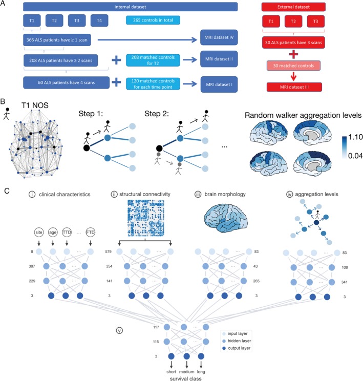
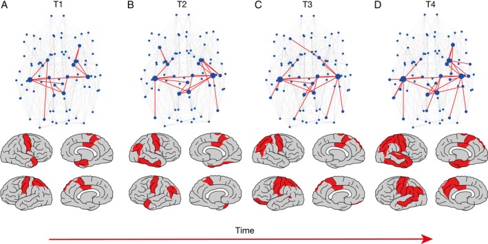
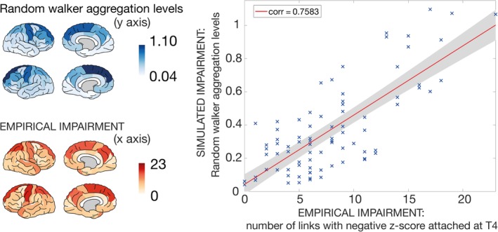
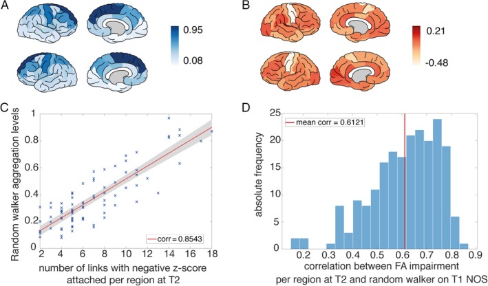
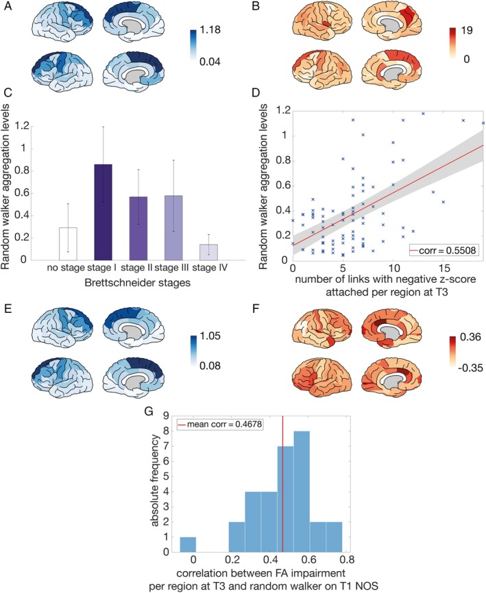
Using brain networks of ALS patients (n = 208) and matched controls across longitudinal time points, network-based statistics unraveled progressive network degeneration originating from the motor cortex and expanding in a spatiotemporal manner. We applied a computational model to the MRI scan of patients to simulate this progressive network degeneration. Simulated aggregation levels at the group and individual level were validated with empirical impairment observed at later time points of white matter and clinical decline using both internal and external datasets.
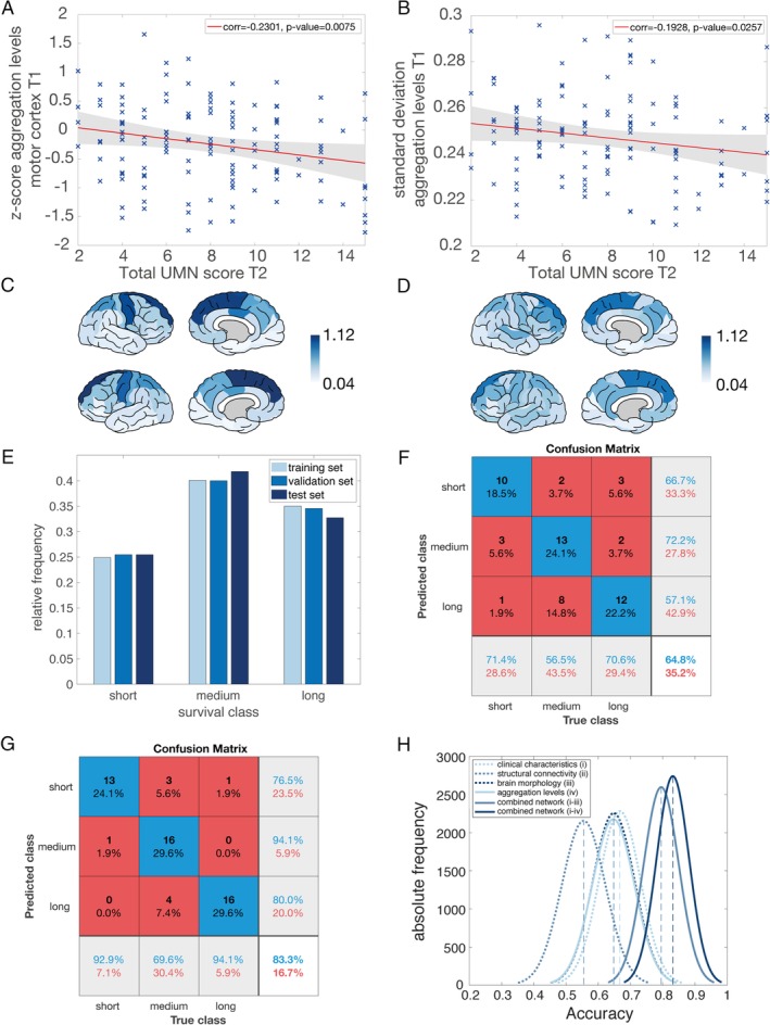
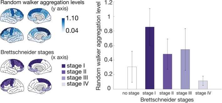
We observe that computer-simulated aggregation levels mimic true disease patterns in ALS patients. Simulated patterns of involvement across cortical areas show significant overlap with the patterns of empirically impaired brain regions on later scans, at both group and individual levels. These findings are validated using an external longitudinal dataset of 30 patients.
Our results are in accordance with established pathological staging systems and may have implications for patient stratification in future clinical trials. Our results demonstrate the utility of computational models in ALS to predict disease progression and underscore their potential as a prognostic biomarker. ANN NEUROL 2020;87:725-738.
尽管出现了定量生物标志物,但肌萎缩侧索硬化症(ALS)的临床试验仍继续依赖于生存或功能量表作为终点。基于神经影像学的 ALS 生物标志物已被证明可在体内检测到与 ALS 相关的病理学,但疾病传播的解剖模式仍描述不足。本研究的目的是使用基于大脑磁共振成像(MRI)数据的网络分析来模拟疾病传播,以预测疾病进展。
通过对 208 名 ALS 患者和匹配对照组的纵向时间点的大脑网络进行分析,网络统计数据揭示了起源于运动皮层并以时空方式扩展的进行性网络退化。我们将计算模型应用于患者的 MRI 扫描中,以模拟这种进行性的网络退化。在个体和组水平上,使用内部和外部数据集,对模拟的 MRI 扫描的白质和临床衰退后的实际损害程度进行了验证。
我们发现计算机模拟的聚集水平可以模拟 ALS 患者的真实疾病模式。模拟的皮层区域受累模式与后期扫描中实际受损脑区的模式在组和个体水平上均有显著重叠。使用 30 名患者的外部纵向数据集验证了这些发现。
我们的研究结果与既定的病理分期系统一致,可能对未来临床试验中的患者分层具有重要意义。我们的结果表明,计算模型在 ALS 中的应用具有预测疾病进展的潜力,并突出了其作为预后生物标志物的潜力。神经病学年鉴 2020;87:725-738.