Bede Peter, Iyer Parameswaran M, Finegan Eoin, Omer Taha, Hardiman Orla
Quantitative Neuroimaging Group, Academic Unit of Neurology, Biomedical Sciences Institute, Trinity College Dublin, Ireland.
Quantitative Neuroimaging Group, Academic Unit of Neurology, Biomedical Sciences Institute, Trinity College Dublin, Ireland.
Neuroimage Clin. 2017 Jun 9;15:653-658. doi: 10.1016/j.nicl.2017.06.010. eCollection 2017.
Diagnostic uncertainty in ALS has serious management implications and delays recruitment into clinical trials. Emerging evidence of presymptomatic disease-burden provides the rationale to develop diagnostic applications based on the evaluation of in-vivo pathological patterns early in the disease.
To outline and test a diagnostic classification approach based on an array of complementary imaging metrics in key disease-associated anatomical structures.
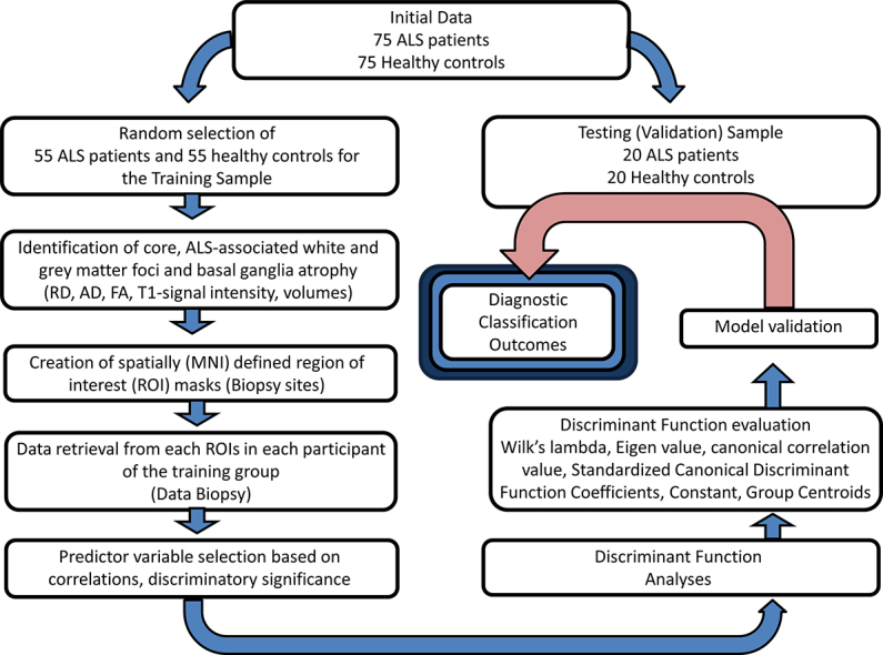
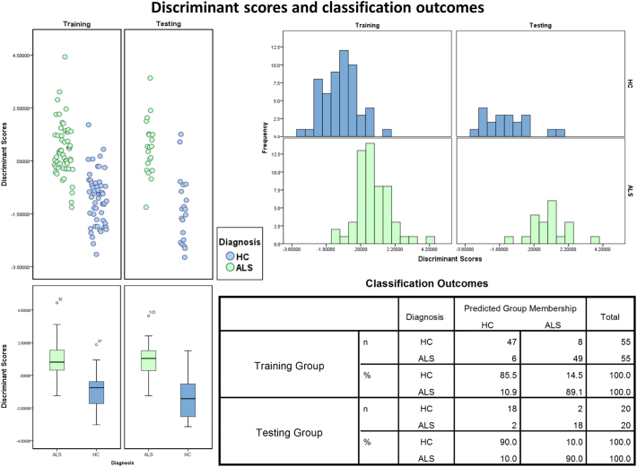
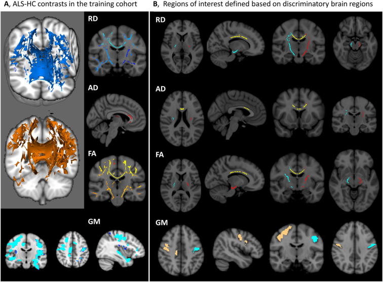
Data from 75 ALS patients and 75 healthy controls were randomly allocated in a 'training' and 'validation' cohort. Spatial masks were created for anatomical foci which best discriminate patients from controls in the 'training sample'. In a virtual 'brain biopsy', data was then retrieved from these key disease-associated brain regions. White matter diffusivity indices, grey matter T1-signal intensity values and basal ganglia volumes were evaluated as predictor variables in a canonical discriminant function.
Following predictor variable selection, a classification specificity of 85.5% and sensitivity of 89.1% was achieved in the training sample and 90% specificity and 90% sensitivity in the validation sample.
This study evaluates disease-associated imaging measures in a dummy diagnostic application. Although larger samples will be required for robust validation, the study confirms the potential of multimodal quantitative imaging in future clinical applications.
肌萎缩侧索硬化症(ALS)诊断的不确定性对治疗管理有着严重影响,并会延迟患者进入临床试验的时间。有症状前疾病负担的新证据为基于疾病早期体内病理模式评估开发诊断应用提供了理论依据。
概述并测试一种基于关键疾病相关解剖结构中一系列互补成像指标的诊断分类方法。
将75例ALS患者和75例健康对照的数据随机分配到“训练”和“验证”队列中。为在“训练样本”中最能区分患者与对照的解剖病灶创建空间掩码。然后在虚拟“脑活检”中,从这些关键疾病相关脑区检索数据。将白质扩散指数、灰质T1信号强度值和基底节体积作为标准判别函数中的预测变量进行评估。
经过预测变量选择,训练样本的分类特异性为85.5%,敏感性为89.1%;验证样本的特异性为90%,敏感性为90%。
本研究在虚拟诊断应用中评估了与疾病相关的成像测量。尽管需要更大的样本进行有力验证,但该研究证实了多模态定量成像在未来临床应用中的潜力。