• 文献检索
  • 文档翻译
  • 深度研究
  • 学术资讯
  • Suppr Zotero 插件Zotero 插件
  • 邀请有礼
  • 套餐&价格
  • 历史记录
应用&插件
Suppr Zotero 插件Zotero 插件浏览器插件Mac 客户端Windows 客户端微信小程序
定价
高级版会员购买积分包购买API积分包
服务
文献检索文档翻译深度研究API 文档MCP 服务
关于我们
关于 Suppr公司介绍联系我们用户协议隐私条款
关注我们

Suppr 超能文献

核心技术专利:CN118964589B侵权必究
粤ICP备2023148730 号-1Suppr @ 2026

文献检索

告别复杂PubMed语法,用中文像聊天一样搜索,搜遍4000万医学文献。AI智能推荐,让科研检索更轻松。

立即免费搜索

文件翻译

保留排版,准确专业,支持PDF/Word/PPT等文件格式,支持 12+语言互译。

免费翻译文档

深度研究

AI帮你快速写综述,25分钟生成高质量综述,智能提取关键信息,辅助科研写作。

立即免费体验

利用光片荧光显微镜研究小鼠视网膜细胞在空间和时间上的行为。

Mouse retinal cell behaviour in space and time using light sheet fluorescence microscopy.

机构信息

Center for Vascular Biology Research and Department of Pathology, Beth Israel Deaconess Medical Center, Harvard Medical School, Boston, United States.

The Beijer Laboratory, Department of Immunology, Genetics and Pathology, Uppsala University, Uppsala, Sweden.

出版信息

Elife. 2020 Feb 19;9:e49779. doi: 10.7554/eLife.49779.

DOI:10.7554/eLife.49779
PMID:32073398
原文链接:https://pmc.ncbi.nlm.nih.gov/articles/PMC7162655/
Abstract

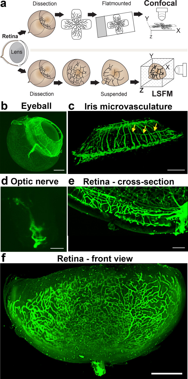
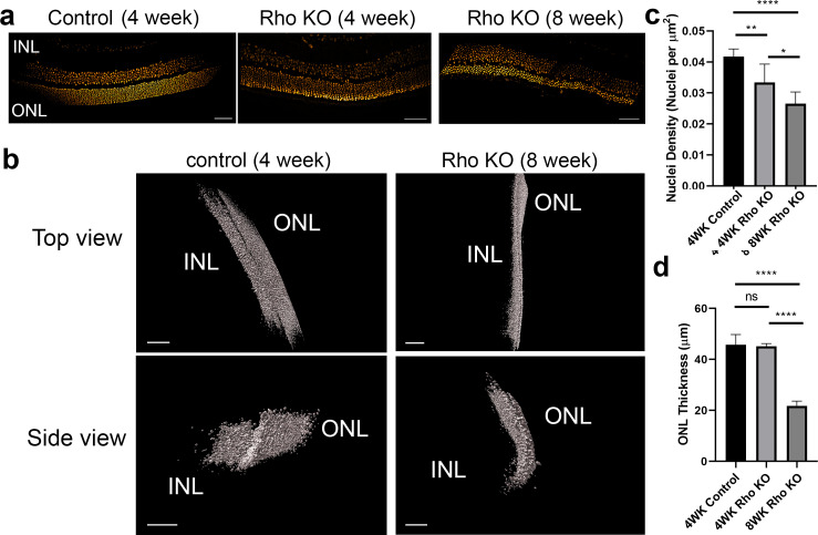
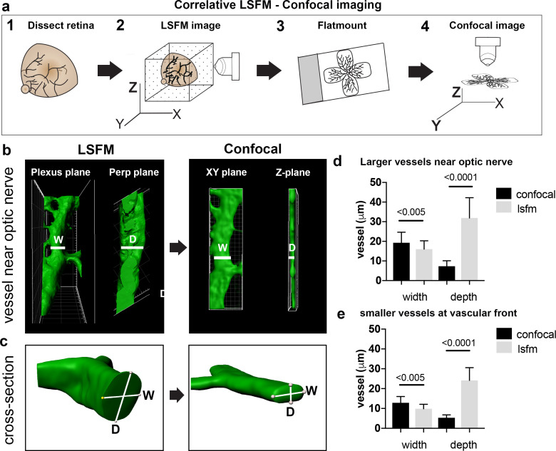
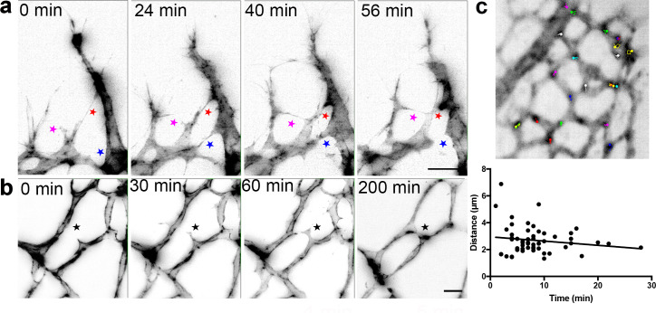
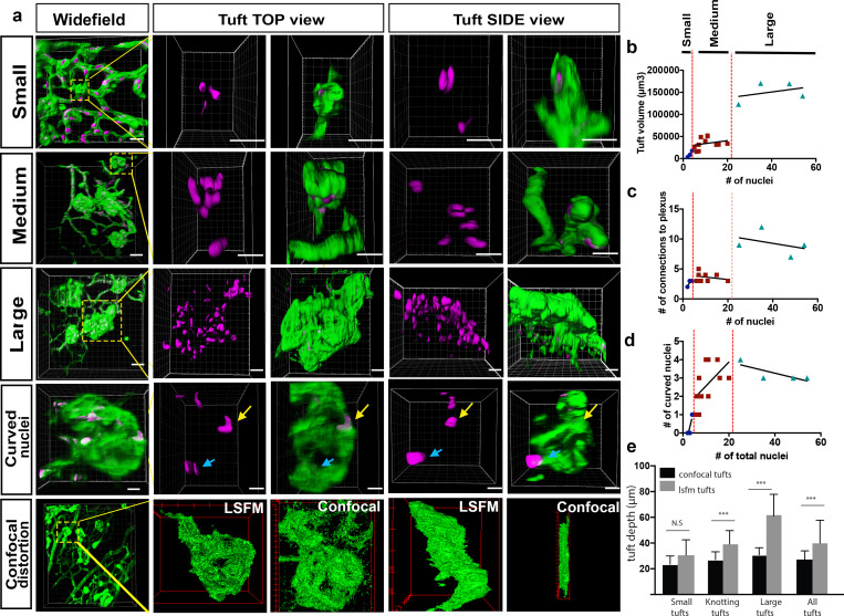
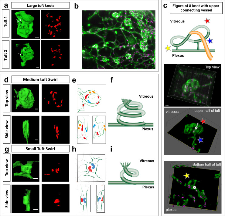
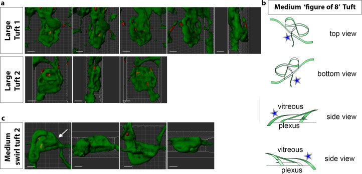
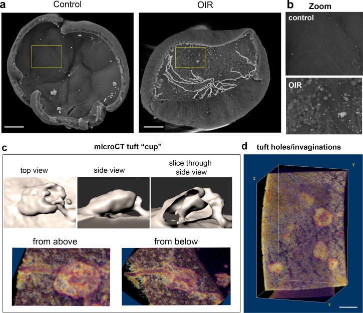
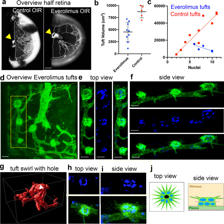
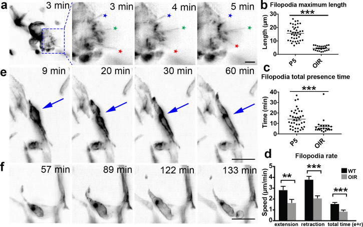
As the general population ages, more people are affected by eye diseases, such as retinopathies. It is therefore critical to improve imaging of eye disease mouse models. Here, we demonstrate that 1) rapid, quantitative 3D and 4D (time lapse) imaging of cellular and subcellular processes in the mouse eye is feasible, with and without tissue clearing, using light-sheet fluorescent microscopy (LSFM); 2) flat-mounting retinas for confocal microscopy significantly distorts tissue morphology, confirmed by quantitative correlative LSFM-Confocal imaging of vessels; 3) LSFM readily reveals new features of even well-studied eye disease mouse models, such as the oxygen-induced retinopathy (OIR) model, including a previously unappreciated 'knotted' morphology to pathological vascular tufts, abnormal cell motility and altered filopodia dynamics when live-imaged. We conclude that quantitative 3D/4D LSFM imaging and analysis has the potential to advance our understanding of the eye, in particular pathological, neurovascular, degenerative processes.

摘要

随着人口老龄化,越来越多的人受到眼病的影响,如视网膜病变。因此,提高眼病小鼠模型的成像质量至关重要。在这里,我们证明了:1)使用光片荧光显微镜(LSFM),无论是否进行组织透明化处理,均可快速、定量地对小鼠眼内的细胞和亚细胞过程进行 3D 和 4D(延时)成像;2)视网膜平铺用于共聚焦显微镜会显著扭曲组织形态,通过对血管进行定量相关的 LSFM-共聚焦成像得到证实;3)LSFM 可以轻松揭示即使是研究得很好的眼病小鼠模型的新特征,例如氧诱导的视网膜病变(OIR)模型,包括以前未被注意到的病理性血管丛的“打结”形态、异常的细胞运动和活细胞成像时丝状伪足动力学的改变。我们得出结论,定量 3D/4D LSFM 成像和分析有可能促进我们对眼睛的理解,特别是病理性、神经血管、退行性过程。

https://cdn.ncbi.nlm.nih.gov/pmc/blobs/8f8c/7162655/dc5584f79c13/elife-49779-resp-fig1.jpg
https://cdn.ncbi.nlm.nih.gov/pmc/blobs/8f8c/7162655/14e00c364cf7/elife-49779-fig1.jpg
https://cdn.ncbi.nlm.nih.gov/pmc/blobs/8f8c/7162655/cbb7a60c231c/elife-49779-fig1-figsupp1.jpg
https://cdn.ncbi.nlm.nih.gov/pmc/blobs/8f8c/7162655/965a01ee6e4a/elife-49779-fig2.jpg
https://cdn.ncbi.nlm.nih.gov/pmc/blobs/8f8c/7162655/40b20a6c378b/elife-49779-fig2-figsupp1.jpg
https://cdn.ncbi.nlm.nih.gov/pmc/blobs/8f8c/7162655/7fe061ce1fd4/elife-49779-fig3.jpg
https://cdn.ncbi.nlm.nih.gov/pmc/blobs/8f8c/7162655/94a5cc187a42/elife-49779-fig4.jpg
https://cdn.ncbi.nlm.nih.gov/pmc/blobs/8f8c/7162655/23745395b75a/elife-49779-fig5.jpg
https://cdn.ncbi.nlm.nih.gov/pmc/blobs/8f8c/7162655/168f659e3ae6/elife-49779-fig6.jpg
https://cdn.ncbi.nlm.nih.gov/pmc/blobs/8f8c/7162655/8c89dba3c122/elife-49779-fig6-figsupp1.jpg
https://cdn.ncbi.nlm.nih.gov/pmc/blobs/8f8c/7162655/819303ab6cc3/elife-49779-fig6-figsupp2.jpg
https://cdn.ncbi.nlm.nih.gov/pmc/blobs/8f8c/7162655/70602c5a4b99/elife-49779-fig6-figsupp3.jpg
https://cdn.ncbi.nlm.nih.gov/pmc/blobs/8f8c/7162655/f658e5d21dc3/elife-49779-fig7.jpg
https://cdn.ncbi.nlm.nih.gov/pmc/blobs/8f8c/7162655/dc5584f79c13/elife-49779-resp-fig1.jpg

相似文献

1
Mouse retinal cell behaviour in space and time using light sheet fluorescence microscopy.利用光片荧光显微镜研究小鼠视网膜细胞在空间和时间上的行为。
Elife. 2020 Feb 19;9:e49779. doi: 10.7554/eLife.49779.
2
Using Light Sheet Fluorescence Microscopy to Image Zebrafish Eye Development.使用光片荧光显微镜对斑马鱼眼睛发育进行成像。
J Vis Exp. 2016 Apr 10(110):e53966. doi: 10.3791/53966.
3
Light-Sheet Microscopy of the Optic Nerve Reveals Axonal Degeneration and Microglial Activation in NMDA-Induced Retinal Injury.视神经的光片显微镜检查揭示了NMDA诱导的视网膜损伤中的轴突退变和小胶质细胞激活。
EC Ophthalmol. 2021 Nov;12(11):23-31. Epub 2021 Oct 28.
4
Three-dimensional Imaging Coupled with Topological Quantification Uncovers Retinal Vascular Plexuses Undergoing Obliteration.三维成像结合拓扑量化揭示正在消失的视网膜血管丛。
Theranostics. 2021 Jan 1;11(3):1162-1175. doi: 10.7150/thno.53073. eCollection 2021.
5
Tissue-culture light sheet fluorescence microscopy (TC-LSFM) allows long-term imaging of three-dimensional cell cultures under controlled conditions.组织培养光片荧光显微镜(TC-LSFM)能够在可控条件下对三维细胞培养物进行长期成像。
Integr Biol (Camb). 2014 Oct;6(10):988-98. doi: 10.1039/c4ib00121d. Epub 2014 Sep 3.
6
Light sheet fluorescence microscopy as a new method for unbiased three-dimensional analysis of vascular injury.光片荧光显微镜作为一种新的无偏三维分析血管损伤的方法。
Cardiovasc Res. 2021 Jan 21;117(2):520-532. doi: 10.1093/cvr/cvaa037.
7
Multiresolution 3D Optical Mapping of Immune Cell Infiltrates in Mouse Asthmatic Lung.多分辨率 3D 光学成像技术在小鼠哮喘肺部中免疫细胞浸润的研究。
Am J Respir Cell Mol Biol. 2023 Jul;69(1):13-21. doi: 10.1165/rcmb.2022-0353MA.
8
Anatomy of the complete mouse eye vasculature explored by light-sheet fluorescence microscopy exposes subvascular-specific remodeling in development and pathology.通过光片荧光显微镜探索的完整小鼠眼脉管系统解剖揭示了发育和病理过程中血管亚特异性重塑。
Exp Eye Res. 2023 Dec;237:109674. doi: 10.1016/j.exer.2023.109674. Epub 2023 Oct 13.
9
Three-dimensional in vivo imaging of the mouse intraocular vasculature during development and disease.小鼠眼内血管系统在发育和疾病过程中的三维体内成像。
Invest Ophthalmol Vis Sci. 2005 Sep;46(9):3021-6. doi: 10.1167/iovs.05-0153.
10
Vascular permeability in retinopathy is regulated by VEGFR2 Y949 signaling to VE-cadherin.血管通透性在视网膜病变中受 VEGFR2 Y949 信号调节至 VE-钙黏蛋白。
Elife. 2020 Apr 21;9:e54056. doi: 10.7554/eLife.54056.

引用本文的文献

1
Association of triglyceride glucose index with diabetic retinopathy in middle-aged and elderly ambulatory type 2 diabetic patients.中老年门诊2型糖尿病患者甘油三酯葡萄糖指数与糖尿病视网膜病变的关联
Front Endocrinol (Lausanne). 2025 Apr 1;16:1442230. doi: 10.3389/fendo.2025.1442230. eCollection 2025.
2
Collagen crosslinking-induced corneal morphological changes: a three-dimensional light sheet Microscopy-based evaluation.胶原交联诱导的角膜形态变化:基于三维光片显微镜的评估。
Sci Rep. 2024 Nov 16;14(1):28330. doi: 10.1038/s41598-024-78516-x.
3
Applications of Lightsheet Fluorescence Microscopy by High Numerical Aperture Detection Lens.

本文引用的文献

1
GNrep mouse: A reporter mouse for front-rear cell polarity.GNrep小鼠:一种用于前后细胞极性的报告基因小鼠。
Genesis. 2019 Jun;57(6):e23299. doi: 10.1002/dvg.23299. Epub 2019 Apr 16.
2
Nuclear rupture at sites of high curvature compromises retention of DNA repair factors.高曲率部位的核破裂会影响 DNA 修复因子的保留。
J Cell Biol. 2018 Nov 5;217(11):3796-3808. doi: 10.1083/jcb.201711161. Epub 2018 Aug 31.
3
Quantifying three-dimensional rodent retina vascular development using optical tissue clearing and light-sheet microscopy.
应用高数值孔径检测透镜的光片荧光显微镜。
J Phys Chem B. 2024 Sep 5;128(35):8273-8289. doi: 10.1021/acs.jpcb.4c01721. Epub 2024 Aug 23.
4
3DVascNet: An Automated Software for Segmentation and Quantification of Mouse Vascular Networks in 3D.3DVascNet:一种用于分割和定量小鼠三维血管网络的自动化软件。
Arterioscler Thromb Vasc Biol. 2024 Jul;44(7):1584-1600. doi: 10.1161/ATVBAHA.124.320672. Epub 2024 May 23.
5
A workflow to visualize vertebrate eyes in 3D.一种可视化三维脊椎动物眼睛的工作流程。
PLoS One. 2023 Aug 22;18(8):e0290420. doi: 10.1371/journal.pone.0290420. eCollection 2023.
6
Expanding the Horizons of Pre-Transplant Renal Vascular Assessment Using Ex Vivo Perfusion.利用体外灌注拓展移植前肾血管评估的视野
Curr Issues Mol Biol. 2023 Jun 29;45(7):5437-5459. doi: 10.3390/cimb45070345.
7
Detection of Choroidal Neovascularization Using Optical Tissue Transparency.利用光学组织透明性检测脉络膜新生血管。
Transl Vis Sci Technol. 2023 Jun 1;12(6):10. doi: 10.1167/tvst.12.6.10.
8
Retinal microvascular and neuronal pathologies probed in vivo by adaptive optical two-photon fluorescence microscopy.自适应光学双光子荧光显微镜在体探测视网膜微血管和神经元病变。
Elife. 2023 Apr 11;12:e84853. doi: 10.7554/eLife.84853.
9
Light-Sheet Microscopy of the Optic Nerve Reveals Axonal Degeneration and Microglial Activation in NMDA-Induced Retinal Injury.视神经的光片显微镜检查揭示了NMDA诱导的视网膜损伤中的轴突退变和小胶质细胞激活。
EC Ophthalmol. 2021 Nov;12(11):23-31. Epub 2021 Oct 28.
10
Endothelial cells during craniofacial development: Populating and patterning the head.颅面发育过程中的内皮细胞:头部的细胞填充与模式形成
Front Bioeng Biotechnol. 2022 Aug 29;10:962040. doi: 10.3389/fbioe.2022.962040. eCollection 2022.
利用光学组织透明化和光片显微镜定量研究三维啮齿动物视网膜血管发育。
J Biomed Opt. 2017 Jul 1;22(7):76011. doi: 10.1117/1.JBO.22.7.076011.
4
Whole-Body Profiling of Cancer Metastasis with Single-Cell Resolution.以单细胞分辨率进行癌症转移的全身分析
Cell Rep. 2017 Jul 5;20(1):236-250. doi: 10.1016/j.celrep.2017.06.010.
5
Mapping of Brain Activity by Automated Volume Analysis of Immediate Early Genes.通过即刻早期基因的自动体积分析绘制大脑活动图
Cell. 2016 Jun 16;165(7):1789-1802. doi: 10.1016/j.cell.2016.05.007. Epub 2016 May 26.
6
Endothelial fluid shear stress sensing in vascular health and disease.血管健康与疾病中的内皮流体剪切应力感知
J Clin Invest. 2016 Mar 1;126(3):821-8. doi: 10.1172/JCI83083.
7
Non-canonical Wnt signalling modulates the endothelial shear stress flow sensor in vascular remodelling.非经典Wnt信号通路在血管重塑过程中调节内皮剪切应力流量传感器。
Elife. 2016 Feb 4;5:e07727. doi: 10.7554/eLife.07727.
8
Simple, Scalable Proteomic Imaging for High-Dimensional Profiling of Intact Systems.用于完整系统高维分析的简单、可扩展蛋白质组学成像
Cell. 2015 Dec 3;163(6):1500-14. doi: 10.1016/j.cell.2015.11.025.
9
Whole-body tissue stabilization and selective extractions via tissue-hydrogel hybrids for high-resolution intact circuit mapping and phenotyping.通过组织-水凝胶杂化体实现全身组织稳定化和选择性提取,用于高分辨率完整回路映射和表型分析。
Nat Protoc. 2015 Nov;10(11):1860-1896. doi: 10.1038/nprot.2015.122. Epub 2015 Oct 22.
10
Clarifying Tissue Clearing.澄清组织透明化
Cell. 2015 Jul 16;162(2):246-257. doi: 10.1016/j.cell.2015.06.067.