Barbacena Pedro, Ouarné Marie, Haigh Jody J, Vasconcelos Francisca F, Pezzarossa Anna, Franco Claudio A
Instituto de Medicina Molecular, Faculdade de Medicina, Universidade de Lisboa, Lisbon, Portugal.
Department of Pharmacology and Therapeutics, Research Institute of Oncology and Hematology, CancerCare Manitoba, University of Manitoba, Winnipeg, Manitoba, Canada.
Genesis. 2019 Jun;57(6):e23299. doi: 10.1002/dvg.23299. Epub 2019 Apr 16.
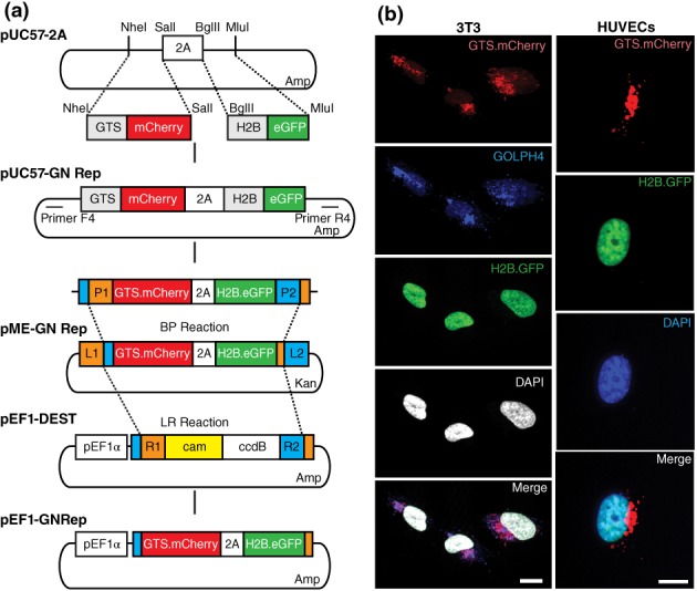
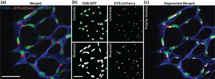
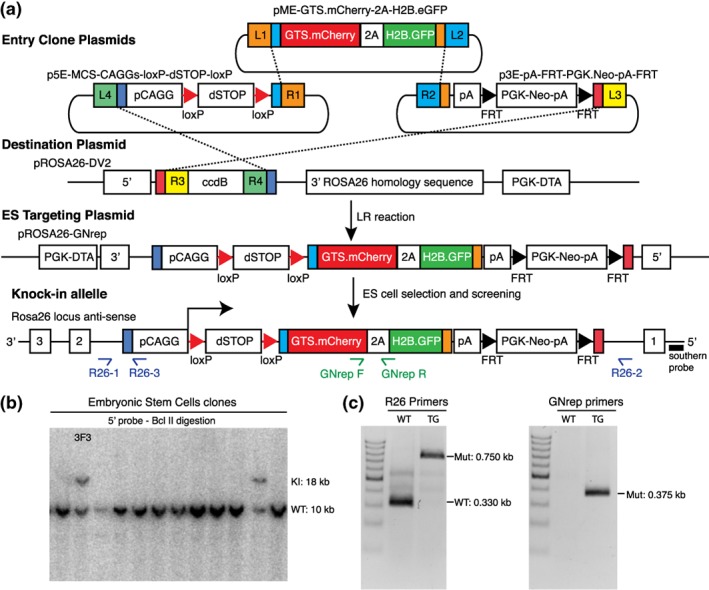
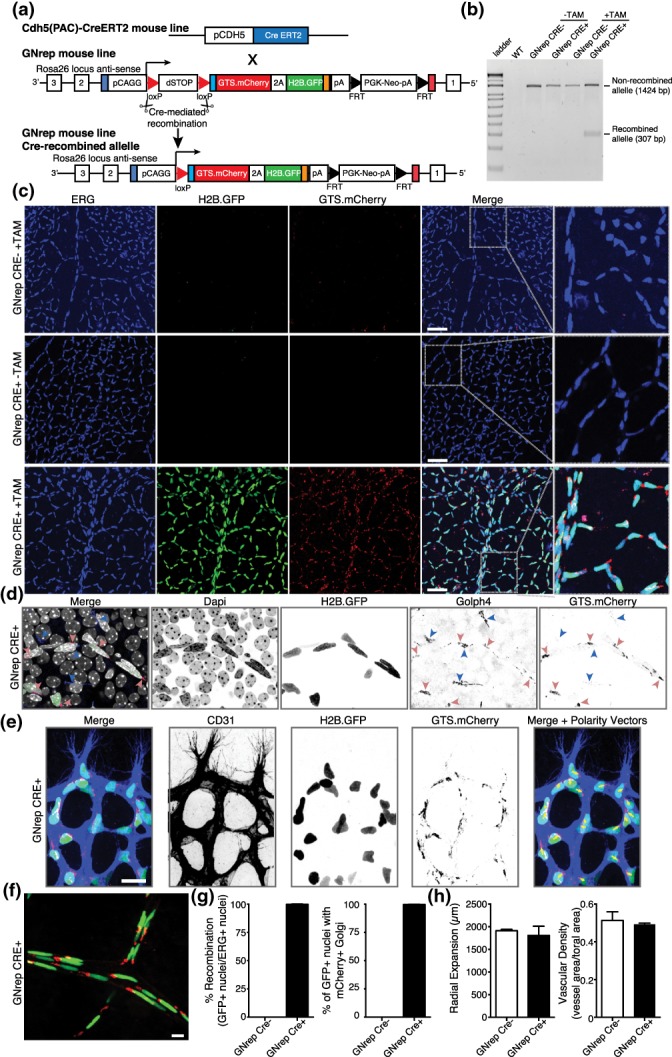
Cell migration is essential during development, regeneration, homeostasis, and disease. Depending on the microenvironment, cells use different mechanisms to migrate. Yet, all modes of migration require the establishment of an intracellular front-rear polarity axis for directional movement. Although front-rear polarity can be easily identified in in vitro conditions, its assessment in vivo by live-imaging is challenging due to tissue complexity and lack of reliable markers. Here, we describe a novel and unique double fluorescent reporter mouse line to study front-rear cell polarity in living tissues, called GNrep. This mouse line simultaneously labels Golgi complexes and nuclei allowing the assignment of a nucleus-to-Golgi axis to each cell, which functions as a readout for cell front-rear polarity. As a proof-of-principle, we validated the efficiency of the GNrep line using an endothelial-specific Cre mouse line. We show that the GNrep labels the nucleus and the Golgi apparatus of endothelial cells with very high efficiency and high specificity. Importantly, the features of fluorescent intensity and localization for both mCherry and eGFP fluorescent intensity and localization allow automated segmentation and assignment of polarity vectors in complex tissues, making GNrep a great tool to study cell behavior in large-scale automated analyses. Altogether, the GNrep mouse line, in combination with different Cre recombinase lines, is a novel and unique tool to study of front-rear polarity in mice, both in fixed tissues or in intravital live imaging. This new line will be instrumental to understand cell migration and polarity in development, homeostasis, and disease.
细胞迁移在发育、再生、体内平衡和疾病过程中至关重要。根据微环境的不同,细胞会采用不同的机制进行迁移。然而,所有迁移模式都需要建立细胞内的前后极性轴以实现定向移动。虽然在体外条件下可以很容易地识别前后极性,但由于组织复杂性和缺乏可靠的标记物,通过活体成像在体内评估它具有挑战性。在这里,我们描述了一种新颖独特的双荧光报告基因小鼠品系,用于研究活组织中的细胞前后极性,称为GNrep。该小鼠品系同时标记高尔基体复合体和细胞核,从而能够为每个细胞确定细胞核到高尔基体的轴,该轴可作为细胞前后极性的一种读数。作为原理验证,我们使用内皮细胞特异性Cre小鼠品系验证了GNrep品系的效率。我们表明,GNrep能够非常高效且高度特异地标记内皮细胞的细胞核和高尔基体。重要的是,mCherry和eGFP荧光强度及定位的特征允许在复杂组织中对极性向量进行自动分割和赋值,这使得GNrep成为在大规模自动分析中研究细胞行为的一个很好的工具。总之,GNrep小鼠品系与不同的Cre重组酶品系相结合,是一种新颖独特的工具,可用于在固定组织或活体成像中研究小鼠的前后极性。这个新的品系将有助于理解发育、体内平衡和疾病过程中的细胞迁移和极性。