Kondiah Pariksha Jolene, Kondiah Pierre P D, Choonara Yahya E, Marimuthu Thashree, Pillay Viness
Wits Advanced Drug Delivery Platform Research Unit, Department of Pharmacy and Pharmacology, School of Therapeutic Sciences, Faculty of Health Sciences, University of the Witwatersrand, Johannesburg, 7 York Road, Parktown 2193, South Africa.
Pharmaceutics. 2020 Feb 17;12(2):166. doi: 10.3390/pharmaceutics12020166.
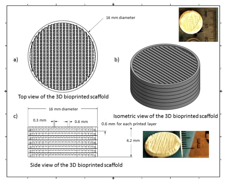
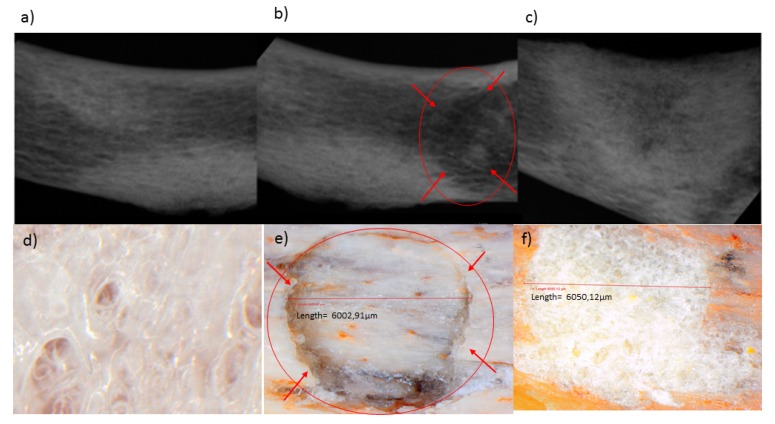
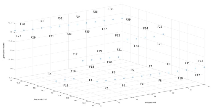
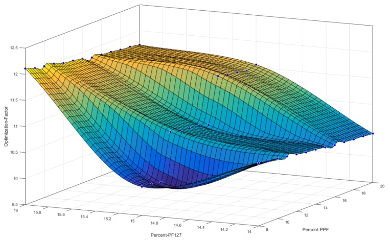
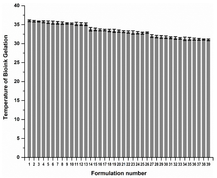
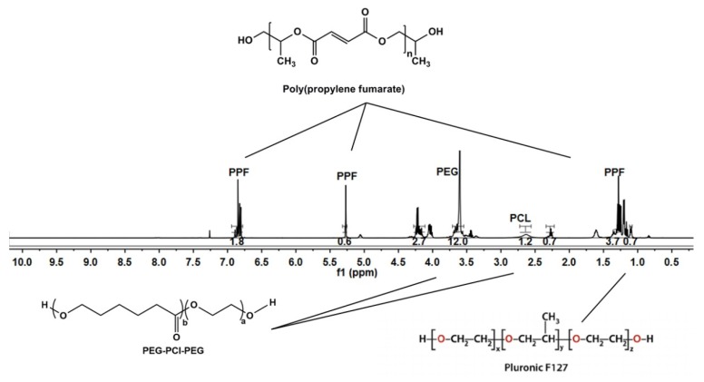
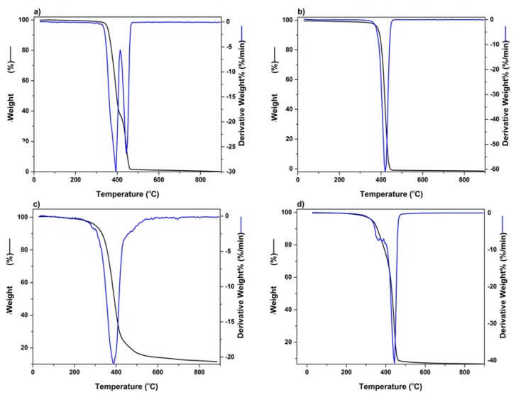
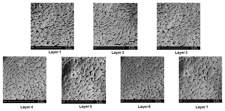
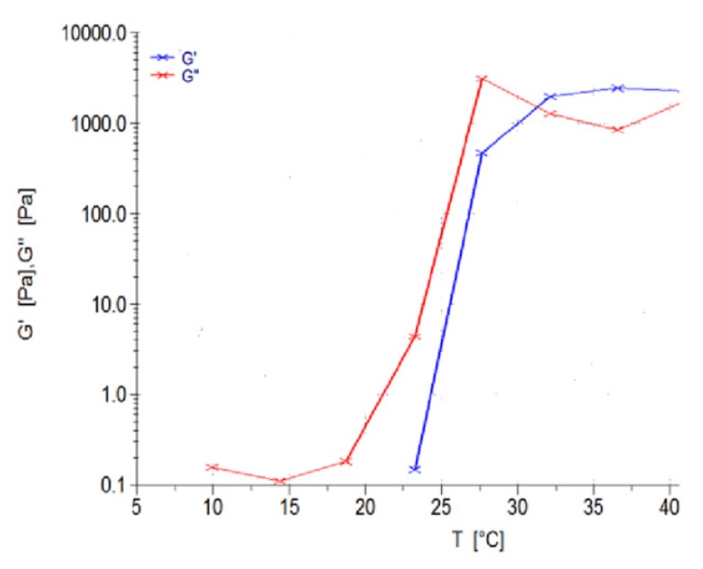
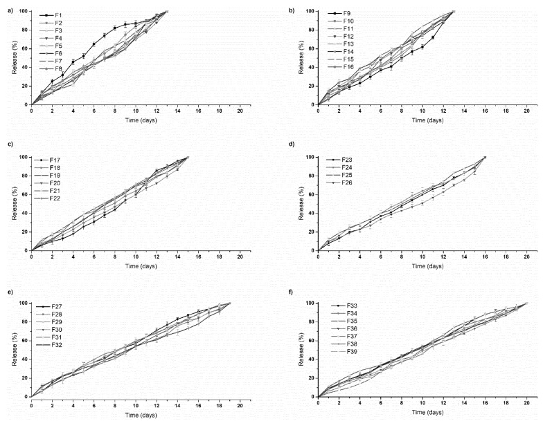
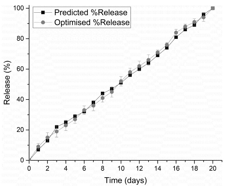
A 3D bioprinted pseudo-bone drug delivery scaffold was fabricated to display matrix strength, matrix resilience, as well as porous morphology of healthy human bone. Computer-aided design (CAD) software was employed for developing the 3D bioprinted scaffold. Further optimization of the scaffold was undertaken using MATLAB software and artificial neural networks (ANN). Polymers employed for formulating the 3D scaffold comprised of polypropylene fumarate (PPF), free radical polymerized polyethylene glycol- polycaprolactone (PEG-PCL-PEG), and pluronic (PF127). Simvastatin was incorporated into the 3D bioprinted scaffolds to further promote bone healing and repair properties. The 3D bioprinted scaffold was characterized for its chemical, morphological, mechanical, and in vitro release kinetics for evaluation of its behavior for application as an implantable scaffold at the site of bone fracture. The ANN-optimized 3D bioprinted scaffold displayed significant properties as a controlled release platform, demonstrating drug release over 20 days. The 3D bioprinted scaffold further displayed formation as a pseudo-bone matrix, using a human clavicle bone model, induced with a butterfly fracture. The strength of the pseudo-bone matrix, evaluated for its matrix hardness (MH) and matrix resilience (MR), was evaluated to be as strong as original bone, having a 99% MH and 98% MR property, to healthy human clavicle bones.
制造了一种3D生物打印的伪骨药物递送支架,以展现健康人骨的基质强度、基质弹性以及多孔形态。采用计算机辅助设计(CAD)软件来开发3D生物打印支架。使用MATLAB软件和人工神经网络(ANN)对支架进行进一步优化。用于配制3D支架的聚合物包括富马酸聚丙烯酯(PPF)、自由基聚合的聚乙二醇-聚己内酯(PEG-PCL-PEG)和普朗尼克(PF127)。将辛伐他汀掺入3D生物打印支架中,以进一步促进骨愈合和修复特性。对3D生物打印支架的化学、形态、力学和体外释放动力学进行表征,以评估其作为骨折部位可植入支架的应用行为。经ANN优化的3D生物打印支架作为控释平台表现出显著特性,在20多天内实现药物释放。使用蝴蝶形骨折诱导的人锁骨骨模型,3D生物打印支架进一步呈现出伪骨基质的形态。对伪骨基质的强度进行评估,其基质硬度(MH)和基质弹性(MR)与健康人锁骨骨相比,分别达到99%的MH和98%的MR,与原始骨一样坚固。