• 文献检索
  • 文档翻译
  • 深度研究
  • 学术资讯
  • Suppr Zotero 插件Zotero 插件
  • 邀请有礼
  • 套餐&价格
  • 历史记录
应用&插件
Suppr Zotero 插件Zotero 插件浏览器插件Mac 客户端Windows 客户端微信小程序
定价
高级版会员购买积分包购买API积分包
服务
文献检索文档翻译深度研究API 文档MCP 服务
关于我们
关于 Suppr公司介绍联系我们用户协议隐私条款
关注我们

Suppr 超能文献

核心技术专利:CN118964589B侵权必究
粤ICP备2023148730 号-1Suppr @ 2026

文献检索

告别复杂PubMed语法,用中文像聊天一样搜索,搜遍4000万医学文献。AI智能推荐,让科研检索更轻松。

立即免费搜索

文件翻译

保留排版,准确专业,支持PDF/Word/PPT等文件格式,支持 12+语言互译。

免费翻译文档

深度研究

AI帮你快速写综述,25分钟生成高质量综述,智能提取关键信息,辅助科研写作。

立即免费体验

NIPA2 通过调节 2 型糖尿病骨质疏松症中的细胞自噬来调节成骨细胞功能。

NIPA2 regulates osteoblast function by modulating mitophagy in type 2 diabetes osteoporosis.

机构信息

Department of Orthopedics, the Fourth Hospital of China Medical University, Shenyang, Liaoning, China.

Department of Orthopedics, the First Hospital of China Medical University, Shenyang, Liaoning, China.

出版信息

Sci Rep. 2020 Feb 20;10(1):3078. doi: 10.1038/s41598-020-59743-4.

DOI:10.1038/s41598-020-59743-4
PMID:32080264
原文链接:https://pmc.ncbi.nlm.nih.gov/articles/PMC7033235/
Abstract

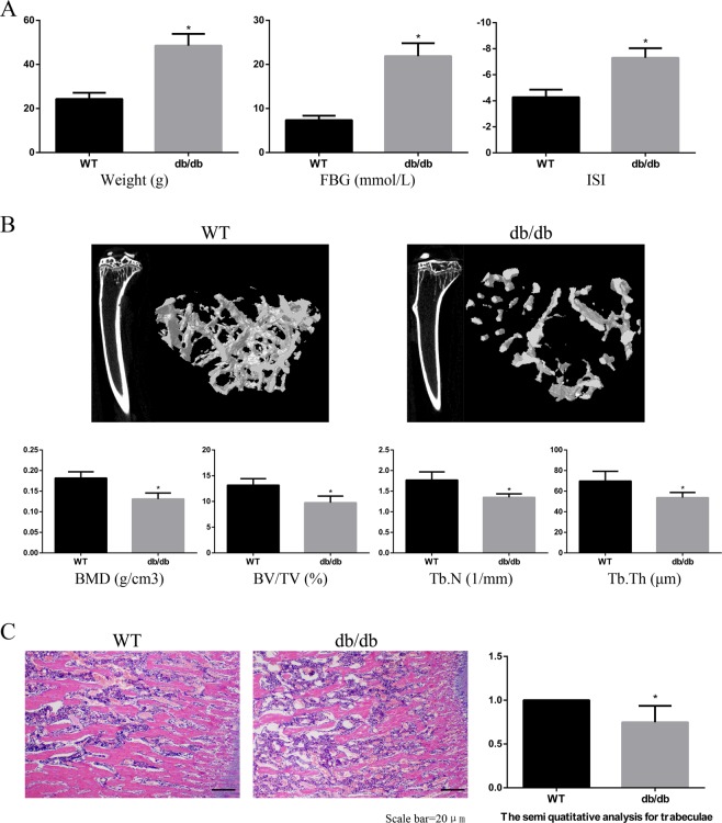
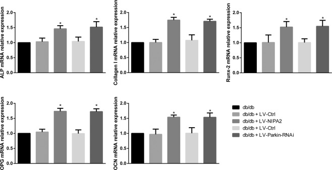
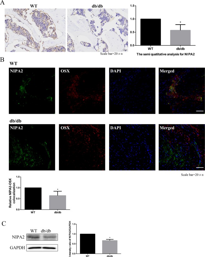
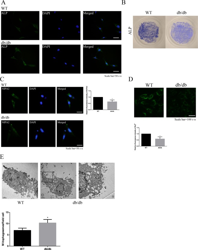
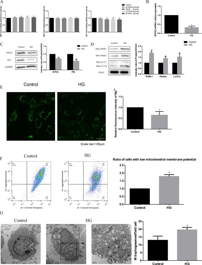
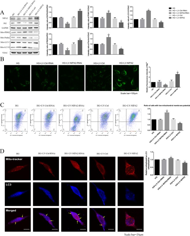
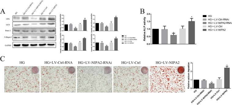
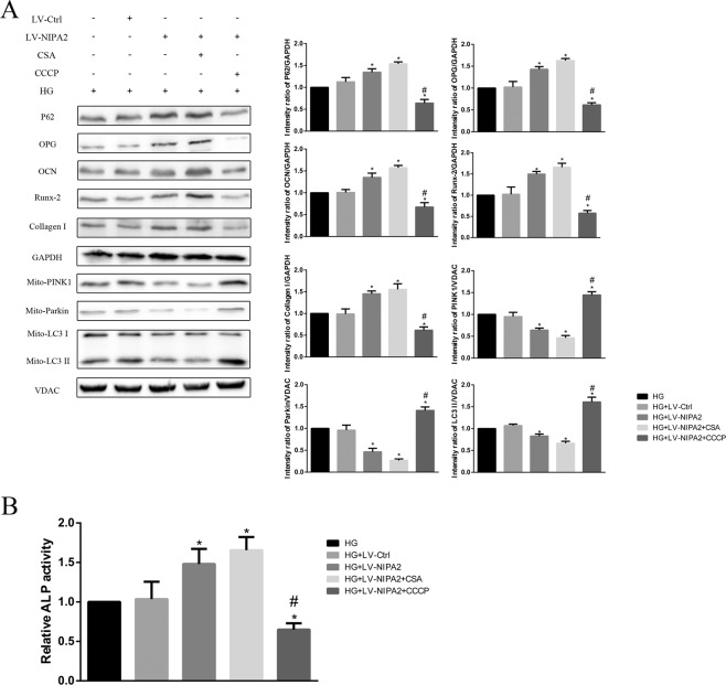
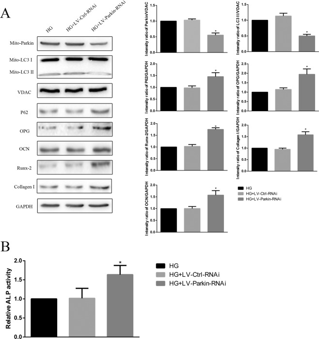
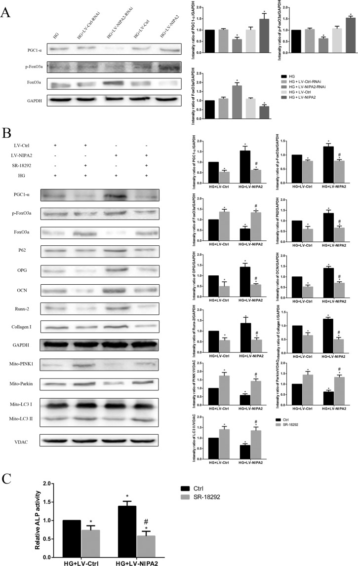
The highly selective magnesium transporter non-imprinted in Prader-Willi/Angelman syndrome region protein 2 (NIPA2) has recently been associated with the development and progression of type 2 diabetes osteoporosis, but the mechanisms involved are still poorly understood. Because mitophagy is involved in the pathology of type 2 diabetes osteoporosis, the present study aimed to explore the relationship among NIPA2, mitophagy and osteoblast osteogenic capacity. NIPA2 expression was reduced in C57BKS background db/db mice and in vitro models of type 2 diabetes osteoporosis, and the activation of mitophagy in primary culture osteoblast-derived from db/db mice and in high glucose-treated human fetal osteoblastic cells (hFOB1.19) was observed. Knockdown, overexpression of NIPA2 and pharmacological inhibition of peroxisome proliferator-activated receptor γ coactivator 1-α (PGC-1α) showed that NIPA2 increased osteoblast function, which was likely regulated by PTEN induced kinase 1 (PINK1)/E3 ubiquitin ligase PARK2 (Parkin)-mediated mitophagy via the PGC-1α/forkhead box O3a(FoxO3a)/mitochondrial membrane potential (MMP) pathway. Furthermore, the negative effect of mitophagy on osteoblast function was confirmed by pharmacological regulation of mitophagy and knockdown of Parkin. Taken together, these results suggest that NIPA2 positively regulates the osteogenic capacity of osteoblasts via the mitophagy pathway in type 2 diabetes.

摘要

高度选择性的镁转运蛋白非印记在 Prader-Willi/Angelman 综合征区域蛋白 2(NIPA2)最近与 2 型糖尿病骨质疏松症的发展和进展有关,但涉及的机制仍知之甚少。由于自噬参与了 2 型糖尿病骨质疏松症的病理学,本研究旨在探讨 NIPA2、自噬和成骨细胞成骨能力之间的关系。在 C57BKS 背景 db/db 小鼠和 2 型糖尿病骨质疏松症的体外模型中,NIPA2 的表达减少,并且在 db/db 小鼠原代培养的成骨细胞和高糖处理的人胎成骨细胞(hFOB1.19)中观察到自噬的激活。NIPA2 的敲低、过表达和过氧化物酶体增殖物激活受体 γ 共激活剂 1-α(PGC-1α)的药理学抑制表明,NIPA2 增加了成骨细胞的功能,这可能是通过 PGC-1α/叉头框 O3a(FoxO3a)/线粒体膜电位(MMP)途径调节的 PTEN 诱导激酶 1(PINK1)/E3 泛素连接酶 PARK2(Parkin)介导的自噬来调节的。此外,通过自噬的药理学调节和 Parkin 的敲低证实了自噬对成骨细胞功能的负向影响。总之,这些结果表明,NIPA2 通过 2 型糖尿病中的自噬途径正向调节成骨细胞的成骨能力。

https://cdn.ncbi.nlm.nih.gov/pmc/blobs/32ad/7033235/a2a285b56c4b/41598_2020_59743_Fig10_HTML.jpg
https://cdn.ncbi.nlm.nih.gov/pmc/blobs/32ad/7033235/62ddac06b620/41598_2020_59743_Fig1_HTML.jpg
https://cdn.ncbi.nlm.nih.gov/pmc/blobs/32ad/7033235/5ccf70cb742d/41598_2020_59743_Fig2_HTML.jpg
https://cdn.ncbi.nlm.nih.gov/pmc/blobs/32ad/7033235/7ca7ba8351d2/41598_2020_59743_Fig3_HTML.jpg
https://cdn.ncbi.nlm.nih.gov/pmc/blobs/32ad/7033235/f2d609e6c24f/41598_2020_59743_Fig4_HTML.jpg
https://cdn.ncbi.nlm.nih.gov/pmc/blobs/32ad/7033235/602d0d8530e3/41598_2020_59743_Fig5_HTML.jpg
https://cdn.ncbi.nlm.nih.gov/pmc/blobs/32ad/7033235/b4ab1ddf4a07/41598_2020_59743_Fig6_HTML.jpg
https://cdn.ncbi.nlm.nih.gov/pmc/blobs/32ad/7033235/62ebcfcfa050/41598_2020_59743_Fig7_HTML.jpg
https://cdn.ncbi.nlm.nih.gov/pmc/blobs/32ad/7033235/8b50b8f9d477/41598_2020_59743_Fig8_HTML.jpg
https://cdn.ncbi.nlm.nih.gov/pmc/blobs/32ad/7033235/82a450d8d6ae/41598_2020_59743_Fig9_HTML.jpg
https://cdn.ncbi.nlm.nih.gov/pmc/blobs/32ad/7033235/a2a285b56c4b/41598_2020_59743_Fig10_HTML.jpg

相似文献

1
NIPA2 regulates osteoblast function by modulating mitophagy in type 2 diabetes osteoporosis.NIPA2 通过调节 2 型糖尿病骨质疏松症中的细胞自噬来调节成骨细胞功能。
Sci Rep. 2020 Feb 20;10(1):3078. doi: 10.1038/s41598-020-59743-4.
2
NIPA2 regulates osteoblast function via its effect on apoptosis pathways in type 2 diabetes osteoporosis.NIPA2 通过影响 2 型糖尿病骨质疏松症中的凋亡途径来调节成骨细胞功能。
Biochem Biophys Res Commun. 2019 Jun 11;513(4):883-890. doi: 10.1016/j.bbrc.2019.04.030. Epub 2019 Apr 16.
3
Local origins impart conserved bone type-related differences in human osteoblast behaviour.局部起源赋予人类成骨细胞行为中与骨类型相关的保守差异。
Eur Cell Mater. 2015 Mar 4;29:155-75; discussion 175-6. doi: 10.22203/ecm.v029a12.
4
Phosphocreatine Promotes Osteoblastic Activities in HO-Induced MC3T3-E1 Cells by Regulating SIRT1/FOXO1/PGC-1α Signaling Pathway.磷酸肌酸通过调节 SIRT1/FOXO1/PGC-1α 信号通路促进 HO 诱导的 MC3T3-E1 细胞成骨活性。
Curr Pharm Biotechnol. 2021;22(5):609-621. doi: 10.2174/1389201021999201116160247.
5
Knockdown of SLC41A1 magnesium transporter promotes mineralization and attenuates magnesium inhibition during osteogenesis of mesenchymal stromal cells.敲低溶质载体家族41成员1(SLC41A1)镁转运体可促进间充质基质细胞成骨过程中的矿化并减弱镁的抑制作用。
Stem Cell Res Ther. 2017 Feb 21;8(1):39. doi: 10.1186/s13287-017-0497-2.
6
Genistein Improves Bone Healing via Triggering Estrogen Receptor Alpha-Mediated Expressions of Osteogenesis-Associated Genes and Consequent Maturation of Osteoblasts.金雀异黄素通过触发雌激素受体 α 介导的成骨相关基因表达及随后的成骨细胞成熟促进骨愈合。
J Agric Food Chem. 2020 Sep 30;68(39):10639-10650. doi: 10.1021/acs.jafc.0c02830. Epub 2020 Sep 8.
7
Insulin-like growth factor-1 promotes osteogenic differentiation and collagen I alpha 2 synthesis via induction of mRNA-binding protein LARP6 expression.胰岛素样生长因子-1通过诱导mRNA结合蛋白LARP6的表达促进成骨分化和I型胶原蛋白α2的合成。
Dev Growth Differ. 2017 Feb;59(2):94-103. doi: 10.1111/dgd.12342. Epub 2017 Feb 17.
8
Peroxisome proliferator-activated receptor activity is involved in the osteoblastic differentiation regulated by bone morphogenetic proteins and tumor necrosis factor-α.过氧化物酶体增殖物激活受体活性参与骨形态发生蛋白和肿瘤坏死因子-α调节的成骨细胞分化。
Mol Cell Endocrinol. 2012 Jan 2;348(1):224-32. doi: 10.1016/j.mce.2011.08.027. Epub 2011 Aug 25.
9
Reduced osteoblast activity in the mice lacking TR4 nuclear receptor leads to osteoporosis.缺乏 TR4 核受体的小鼠成骨细胞活性降低导致骨质疏松症。
Reprod Biol Endocrinol. 2012 Jun 7;10:43. doi: 10.1186/1477-7827-10-43.
10
Advanced glycation end products affect growth and function of osteoblasts.糖基化终产物影响成骨细胞的生长和功能。
Clin Exp Rheumatol. 2011 Jul-Aug;29(4):650-60. Epub 2011 Aug 31.

引用本文的文献

1
Different Forms of Regulated Cell Death in Type-2-Diabetes-Mellitus-Related Osteoporosis: A Focus on Mechanisms and Therapeutic Strategies.2型糖尿病相关骨质疏松症中不同形式的程序性细胞死亡:聚焦机制与治疗策略
Int J Mol Sci. 2025 May 6;26(9):4417. doi: 10.3390/ijms26094417.
2
The Roles of Forkhead Box O3a (FOXO3a) in Bone and Cartilage Diseases - A Narrative Review.叉头框O3a(FOXO3a)在骨与软骨疾病中的作用——一篇叙述性综述
Drug Des Devel Ther. 2025 Feb 27;19:1357-1375. doi: 10.2147/DDDT.S494841. eCollection 2025.
3
Enhanced SIRT3 expression restores mitochondrial quality control mechanism to reverse osteogenic impairment in type 2 diabetes mellitus.

本文引用的文献

1
UBE2C promotes the progression of head and neck squamous cell carcinoma.UBE2C 促进头颈部鳞状细胞癌的进展。
Biochem Biophys Res Commun. 2020 Mar 5;523(2):389-397. doi: 10.1016/j.bbrc.2019.12.064. Epub 2019 Dec 20.
2
Responses of primary osteoblasts and osteoclasts from hemizygous β-globin knockout thalassemic mice with elevated plasma glucose to 1,25-dihydroxyvitamin D.杂合β-珠蛋白基因敲除地中海贫血小鼠高血糖状态下原代成骨细胞和破骨细胞对 1,25-二羟维生素 D 的反应。
Sci Rep. 2019 Sep 27;9(1):13963. doi: 10.1038/s41598-019-50414-7.
3
Deficiency of stress-associated gene increases bone volume by attenuating differentiation of osteoclasts and enhancing differentiation of osteoblasts.
增强的SIRT3表达可恢复线粒体质量控制机制,以逆转2型糖尿病中的成骨障碍。
Bone Res. 2025 Mar 3;13(1):30. doi: 10.1038/s41413-024-00399-5.
4
Clinical Implications of Circulating Tumor DNA in Multiple Myeloma and Its Precursor Diseases.循环肿瘤DNA在多发性骨髓瘤及其前驱疾病中的临床意义
Ann Lab Med. 2025 May 1;45(3):279-290. doi: 10.3343/alm.2024.0424. Epub 2025 Feb 28.
5
Molecular insights into the interplay between type 2 diabetes mellitus and osteoporosis: implications for endocrine health.2型糖尿病与骨质疏松症相互作用的分子机制:对内分泌健康的影响
Front Endocrinol (Lausanne). 2025 Jan 17;15:1483512. doi: 10.3389/fendo.2024.1483512. eCollection 2024.
6
Bioinformatic approach to explain how Mg from seawater may be incorporated into coral skeletons.用生物信息学方法解释海水中的镁如何被纳入珊瑚骨骼。
R Soc Open Sci. 2025 Jan 22;12(1):232011. doi: 10.1098/rsos.232011. eCollection 2025 Jan.
7
The inhibition of PINK1/Drp1-mediated mitophagy by hyperglycemia leads to impaired osteoblastogenesis in diabetes.高血糖对PINK1/Drp1介导的线粒体自噬的抑制作用会导致糖尿病中骨生成受损。
iScience. 2024 Dec 3;28(1):111519. doi: 10.1016/j.isci.2024.111519. eCollection 2025 Jan 17.
8
extract regulates oxidative stress to maintain calcium homeostasis and improve diabetic osteoporosis.提取物可调节氧化应激,以维持钙稳态并改善糖尿病性骨质疏松症。
Food Sci Nutr. 2024 Aug 20;12(10):8067-8083. doi: 10.1002/fsn3.4413. eCollection 2024 Oct.
9
is positively correlated with in infection and a biomarker for mucosal injury.与感染呈正相关,是黏膜损伤的生物标志物。
iScience. 2024 Jun 20;27(7):110323. doi: 10.1016/j.isci.2024.110323. eCollection 2024 Jul 19.
10
Long non-coding RNA MIAT serves as a biomarker of fragility fracture and promotes fracture healing.长链非编码 RNA MIAT 可作为脆性骨折的生物标志物,并促进骨折愈合。
J Orthop Surg Res. 2024 Jun 8;19(1):343. doi: 10.1186/s13018-024-04824-7.
应激相关基因缺失通过减弱破骨细胞分化和增强成骨细胞分化来增加骨量。
FASEB J. 2019 Aug;33(8):8836-8852. doi: 10.1096/fj.201802322RR. Epub 2019 May 8.
4
Effect of magnesium on reducing the UV-induced oxidative damage in marrow mesenchymal stem cells.镁对减少骨髓间充质干细胞中 UV 诱导的氧化损伤的影响。
J Biomed Mater Res A. 2019 Jun;107(6):1253-1263. doi: 10.1002/jbm.a.36634. Epub 2019 Feb 15.
5
Osteoporosis of the vertebra and osteochondral remodeling of the endplate causes intervertebral disc degeneration in ovariectomized mice.去卵巢小鼠的椎体骨质疏松症和终板骨软骨重塑导致椎间盘退变。
Arthritis Res Ther. 2018 Sep 10;20(1):207. doi: 10.1186/s13075-018-1701-1.
6
NR4A1 Promotes Diabetic Nephropathy by Activating Mff-Mediated Mitochondrial Fission and Suppressing Parkin-Mediated Mitophagy.NR4A1通过激活Mff介导的线粒体分裂和抑制Parkin介导的线粒体自噬促进糖尿病肾病。
Cell Physiol Biochem. 2018;48(4):1675-1693. doi: 10.1159/000492292. Epub 2018 Aug 3.
7
Effects of Type 2 Diabetic Serum on Proliferation and Osteogenic Differentiation of Mesenchymal Stem Cells.2 型糖尿病血清对间充质干细胞增殖及成骨分化的影响。
J Diabetes Res. 2018 Jun 5;2018:5765478. doi: 10.1155/2018/5765478. eCollection 2018.
8
Defective autophagy in osteoblasts induces endoplasmic reticulum stress and causes remarkable bone loss.成骨细胞中的自噬缺陷会诱导内质网应激,导致显著的骨丢失。
Autophagy. 2018;14(10):1726-1741. doi: 10.1080/15548627.2018.1483807. Epub 2018 Jul 28.
9
Magnesium and Human Health: Perspectives and Research Directions.镁与人类健康:观点与研究方向
Int J Endocrinol. 2018 Apr 16;2018:9041694. doi: 10.1155/2018/9041694. eCollection 2018.
10
Irbesartan attenuates advanced glycation end products-mediated damage in diabetes-associated osteoporosis through the AGEs/RAGE pathway.厄贝沙坦通过 AGEs/RAGE 途径减轻糖尿病相关骨质疏松症中晚期糖基化终产物介导的损伤。
Life Sci. 2018 Jul 15;205:184-192. doi: 10.1016/j.lfs.2018.04.042. Epub 2018 Apr 24.