Wang Min, Liao Haiyan, Shen Qin, Cai Sainan, Zhang Hongchun, Xiang Yijuan, Liu Siyu, Wang Tianyu, Zi Yuheng, Mao Zhenni, Tan Changlian
Department of Radiology, The Second Xiangya Hospital, Central South University, Changsha, China.
Department of Radiology, The First Affiliated Hospital, University of Science and Technology of China, Changsha, China.
Front Neurol. 2020 Jan 31;11:28. doi: 10.3389/fneur.2020.00028. eCollection 2020.
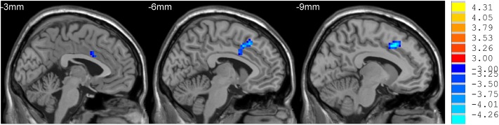
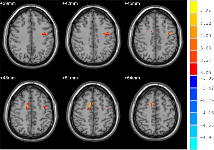
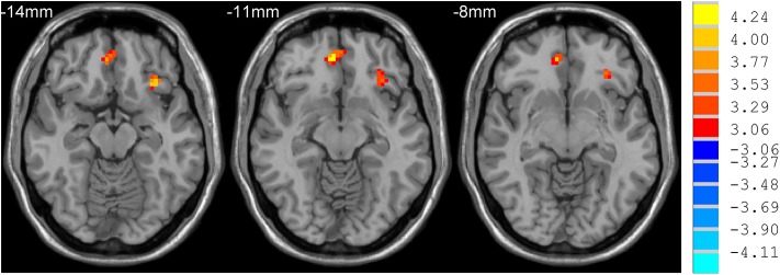
Depression is reported to occur 5-10 years early than the onset of motor symptoms in Parkinson (PD) patients. However, markers for early diagnosis of PD in individuals with sub-clinical depression still remain to be identified. This study utilized Regional Homogeneity (ReHo) to investigate the alterations in resting state brain activities in Parkinson (PD) patients with different degrees of depression. Twenty non-depressed PD patients, twenty mild to moderately depressed PD patients, and thirteen severely depressed PD patients were recruited. Hamilton Depression Scale (HDS) and the Beck Depression Inventory (BDI) were assessed depression. Resting-state functional magnetic resonance imaging (rs-MRI) was analyzed with ReHo. PD patients with mild to moderate depression had decreased ReHo in the left dorsal anterior cingulate cortex when compared with PD patients without depression. PD patients with severe depression exhibited increased ReHo in the left inferior prefrontal gyrus and right orbitofrontal area when compared with PD patients with mild to moderate depression. ReHo values in the bilateral supplementary motor area (SMA) in PD patients with severe depression was also increased when compared with PD patients without depression. This study suggests that rs-MRI with ReHo analysis can detect early changes in brain function that associate with depression in PD patients, which could be biomarkers for early diagnosis and treatment of PD related depression.
据报道,抑郁症在帕金森病(PD)患者中比运动症状出现早5至10年。然而,亚临床抑郁症个体中PD早期诊断的标志物仍有待确定。本研究利用局部一致性(ReHo)来研究不同抑郁程度的帕金森病(PD)患者静息态脑活动的改变。招募了20名无抑郁的PD患者、20名轻度至中度抑郁的PD患者和13名重度抑郁的PD患者。采用汉密尔顿抑郁量表(HDS)和贝克抑郁量表(BDI)评估抑郁情况。用ReHo分析静息态功能磁共振成像(rs-MRI)。与无抑郁的PD患者相比,轻度至中度抑郁的PD患者左侧背侧前扣带回皮质的ReHo降低。与轻度至中度抑郁的PD患者相比,重度抑郁的PD患者左侧额下回和右侧眶额区的ReHo增加。与无抑郁的PD患者相比,重度抑郁的PD患者双侧辅助运动区(SMA)的ReHo值也增加。本研究表明,采用ReHo分析的rs-MRI可以检测出与PD患者抑郁相关的脑功能早期变化,这可能是PD相关抑郁早期诊断和治疗的生物标志物。