• 文献检索
  • 文档翻译
  • 深度研究
  • 学术资讯
  • Suppr Zotero 插件Zotero 插件
  • 邀请有礼
  • 套餐&价格
  • 历史记录
应用&插件
Suppr Zotero 插件Zotero 插件浏览器插件Mac 客户端Windows 客户端微信小程序
定价
高级版会员购买积分包购买API积分包
服务
文献检索文档翻译深度研究API 文档MCP 服务
关于我们
关于 Suppr公司介绍联系我们用户协议隐私条款
关注我们

Suppr 超能文献

核心技术专利:CN118964589B侵权必究
粤ICP备2023148730 号-1Suppr @ 2026

文献检索

告别复杂PubMed语法,用中文像聊天一样搜索,搜遍4000万医学文献。AI智能推荐,让科研检索更轻松。

立即免费搜索

文件翻译

保留排版,准确专业,支持PDF/Word/PPT等文件格式,支持 12+语言互译。

免费翻译文档

深度研究

AI帮你快速写综述,25分钟生成高质量综述,智能提取关键信息,辅助科研写作。

立即免费体验

准分子激光原位角膜磨镶术(LASIK)瓣厚度对角膜生物力学行为的影响:有限元分析

Effects of the LASIK flap thickness on corneal biomechanical behavior: a finite element analysis.

作者信息

Fang Lihua, Wang Yan, Yang Ruizhi, Deng Sijing, Deng Jiahao, Wan Linsun

机构信息

Key Laboratory of National Engineering Laboratory for Nondestructive Testing and Optoelectric Sensing Technology and Application (Ministry of Education), Nanchang Hangkong University, Add: No 696. Fenghenan Rd, Donghu District, Nanchang city, Jiangxi Province, 330063, China.

Tianjin Eye Hospital & Eye Institute, Ophthalmology and Visual Development Key Laboratory, Tianjin Medical University, Tianjin, 300020, China.

出版信息

BMC Ophthalmol. 2020 Feb 24;20(1):67. doi: 10.1186/s12886-020-01338-8.

DOI:10.1186/s12886-020-01338-8
PMID:32093676
原文链接:https://pmc.ncbi.nlm.nih.gov/articles/PMC7038569/
Abstract

BACKGROUND

It is well known that the biomechanical properties change after LASIK refractive surgery. One reason is the impact of flap creation on the residual stroma. The results have revealed that the change is closely related with the flap thickness in several studies. However, the quantitative relationships between the distributions of displacement and stress on the corneal surface and flap thickness have not been studied. The aim of the study was to quantify evaluate the biomechanical change caused by the LASIK flap.

METHODS

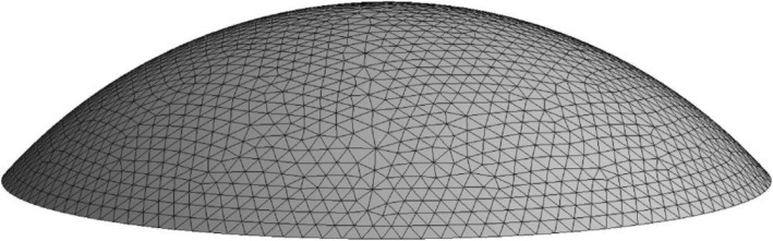
By building a finite element model of the cornea, the displacement, the stress and the strain on the corneal surface were analyzed.

RESULTS

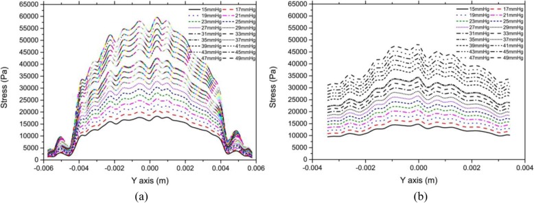
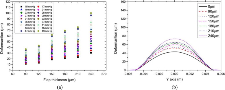
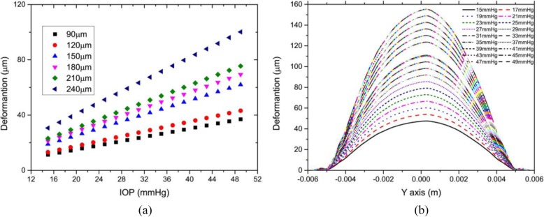
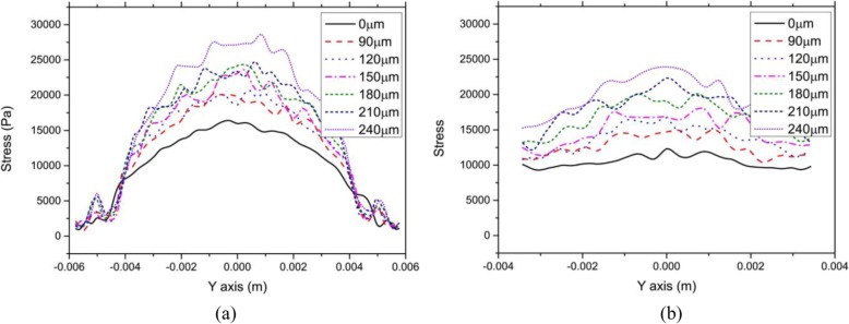
The results showed that the corneal flap could obviously cause the deformation of the anterior corneal surface. For example, the displacement of the corneal vertex achieved 15 μm more than that without corneal flap, when the thickness of corneal flap was 120 μm thick. This displacement was enough to cause the change of aberrations in the human eyes. In the central part of the cornea, the stress on the anterior corneal surface increased with flap thickness. But the change in the stress on the posterior corneal surface was significantly less than that on the anterior surface. In addition, the stress in the central part of the anterior corneal surface increased significantly as the intra-ocular pressure (IOP) increase. Furthermore the increase of IOP had a clearly less effect on stress distribution at the edge of the cornea. Distributions of strain on the corneal surface were similar to those of stress.

CONCLUSIONS

The changes in the biomechanical properties of cornea after refractive surgery should not be ignored.

摘要

背景

众所周知,准分子激光原位角膜磨镶术(LASIK)屈光手术后生物力学特性会发生改变。其中一个原因是角膜瓣制作对剩余基质的影响。多项研究结果表明,这种改变与角膜瓣厚度密切相关。然而,角膜表面位移和应力分布与角膜瓣厚度之间的定量关系尚未得到研究。本研究的目的是定量评估LASIK角膜瓣引起的生物力学变化。

方法

通过构建角膜的有限元模型,分析角膜表面的位移、应力和应变。

结果

结果显示,角膜瓣可明显导致角膜前表面变形。例如,当角膜瓣厚度为120μm时,角膜顶点的位移比无角膜瓣时多15μm。这种位移足以引起人眼像差的变化。在角膜中央部分,角膜前表面的应力随角膜瓣厚度增加而增大。但角膜后表面应力的变化明显小于前表面。此外,随着眼内压(IOP)升高,角膜前表面中央部分的应力显著增加。而且,眼内压升高对角膜边缘应力分布的影响明显较小。角膜表面应变分布与应力分布相似。

结论

屈光手术后角膜生物力学特性的变化不容忽视。

https://cdn.ncbi.nlm.nih.gov/pmc/blobs/f90d/7038569/dfc54f1d5566/12886_2020_1338_Fig7_HTML.jpg
https://cdn.ncbi.nlm.nih.gov/pmc/blobs/f90d/7038569/4f59597267c6/12886_2020_1338_Fig1_HTML.jpg
https://cdn.ncbi.nlm.nih.gov/pmc/blobs/f90d/7038569/da606727f1e5/12886_2020_1338_Fig2_HTML.jpg
https://cdn.ncbi.nlm.nih.gov/pmc/blobs/f90d/7038569/20cc1b671d03/12886_2020_1338_Fig3_HTML.jpg
https://cdn.ncbi.nlm.nih.gov/pmc/blobs/f90d/7038569/a5a78e179552/12886_2020_1338_Fig4_HTML.jpg
https://cdn.ncbi.nlm.nih.gov/pmc/blobs/f90d/7038569/b07454a60e5e/12886_2020_1338_Fig5_HTML.jpg
https://cdn.ncbi.nlm.nih.gov/pmc/blobs/f90d/7038569/ffd9d04a1b82/12886_2020_1338_Fig6_HTML.jpg
https://cdn.ncbi.nlm.nih.gov/pmc/blobs/f90d/7038569/dfc54f1d5566/12886_2020_1338_Fig7_HTML.jpg

相似文献

1
Effects of the LASIK flap thickness on corneal biomechanical behavior: a finite element analysis.准分子激光原位角膜磨镶术(LASIK)瓣厚度对角膜生物力学行为的影响:有限元分析
BMC Ophthalmol. 2020 Feb 24;20(1):67. doi: 10.1186/s12886-020-01338-8.
2
Changes in biomechanical properties of the cornea and intraocular pressure after myopic laser in situ keratomileusis using a femtosecond laser for flap creation determined using ocular response analyzer and Goldmann applanation tonometry.应用飞秒激光制作角膜瓣的近视激光原位角膜磨镶术后,应用眼反应分析仪和压平眼压计检测角膜生物力学特性和眼压的变化。
J Glaucoma. 2015 Mar;24(3):195-201. doi: 10.1097/IJG.0b013e31829da1ec.
3
Effects of altered corneal stiffness on native and postoperative LASIK corneal biomechanical behavior: A whole-eye finite element analysis.角膜硬度改变对原位及准分子激光原位角膜磨镶术后角膜生物力学行为的影响:全眼有限元分析
J Refract Surg. 2009 Oct;25(10):875-87. doi: 10.3928/1081597X-20090917-09. Epub 2009 Oct 12.
4
Changes in ocular biomechanics after femtosecond laser creation of a laser in situ keratomileusis flap.飞秒激光制作准分子原位角膜磨镶术瓣后眼生物力学的变化。
J Cataract Refract Surg. 2016 Jan;42(1):127-31. doi: 10.1016/j.jcrs.2015.07.047.
5
Comparison of biomechanical effects of small-incision lenticule extraction and laser in situ keratomileusis: finite-element analysis.小切口透镜切除术与准分子原位角膜磨镶术生物力学效应的比较:有限元分析
J Cataract Refract Surg. 2014 Jun;40(6):971-80. doi: 10.1016/j.jcrs.2013.08.065.
6
Randomized, contralateral eye study to evaluate the effect of standard and inverted side-cut angle on corneal biomechanical properties during femtosecond laser-assisted in situ keratomileusis.随机、对侧眼研究评估飞秒激光辅助原位角膜磨镶术中标准和倒转侧切口角度对角膜生物力学特性的影响。
Acta Ophthalmol. 2014 Sep;92(6):e437-42. doi: 10.1111/aos.12396. Epub 2014 Mar 25.
7
Refractive change induced by the LASIK flap in a biomechanical finite element model.
J Refract Surg. 2006 Mar;22(3):286-92. doi: 10.3928/1081-597X-20060301-15.
8
Measurement of corneal curvature change after mechanical laser in situ keratomileusis flap creation and femtosecond laser flap creation.机械法准分子原位角膜磨镶术(LASIK)制作角膜瓣和飞秒激光制作角膜瓣后角膜曲率变化的测量。
J Cataract Refract Surg. 2008 Feb;34(2):238-42. doi: 10.1016/j.jcrs.2007.09.023.
9
Postoperative changes in intraocular pressure and corneal biomechanical metrics Laser in situ keratomileusis versus laser-assisted subepithelial keratectomy.准分子原位角膜磨镶术与准分子激光上皮瓣下角膜磨镶术后眼压及角膜生物力学指标的变化
J Cataract Refract Surg. 2009 Oct;35(10):1774-88. doi: 10.1016/j.jcrs.2009.05.041.
10
Theoretical Analysis of Wave-Front Aberrations Induced from Conventional Laser Refractive Surgery in a Biomechanical Finite Element Model.基于生物力学有限元模型的常规激光屈光手术对波前像差的理论分析。
Invest Ophthalmol Vis Sci. 2020 May 11;61(5):34. doi: 10.1167/iovs.61.5.34.

引用本文的文献

1
A review of human cornea finite element modeling: geometry modeling, constitutive modeling, and outlooks.人眼角膜有限元建模综述:几何建模、本构建模及展望
Front Bioeng Biotechnol. 2024 Oct 15;12:1455027. doi: 10.3389/fbioe.2024.1455027. eCollection 2024.
2
Study of corneal and retinal thicknesses at five years after FS-LASIK and SMILE for myopia.FS-LASIK 和 SMILE 治疗近视五年后角膜和视网膜厚度的研究。
BMC Ophthalmol. 2024 Sep 5;24(1):396. doi: 10.1186/s12886-024-03661-w.
3
Ex vivo, in vivo and in silico studies of corneal biomechanics: a systematic review.

本文引用的文献

1
Optimization of surgical parameters based on patient-specific models: Application to arcuate keratotomy.基于患者特定模型的手术参数优化:在弧形角膜切开术中的应用。
J Cataract Refract Surg. 2019 Aug;45(8):1084-1091. doi: 10.1016/j.jcrs.2019.02.022.
2
Tensile biomechanical properties and constitutive parameters of human corneal stroma extracted by SMILE procedure.经 SMILE 手术提取的人眼角膜基质的拉伸生物力学特性和本构参数。
J Mech Behav Biomed Mater. 2018 Sep;85:102-108. doi: 10.1016/j.jmbbm.2018.05.042. Epub 2018 May 30.
3
Biomechanical Changes After LASIK Flap Creation Combined With Rapid Cross-Linking Measured With Brillouin Microscopy.
角膜生物力学的离体、在体和计算研究:系统评价。
Phys Eng Sci Med. 2024 Jun;47(2):403-441. doi: 10.1007/s13246-024-01403-2. Epub 2024 Apr 10.
4
Study of the Influence of Boundary Conditions on Corneal Deformation Based on the Finite Element Method of a Corneal Biomechanics Model.基于角膜生物力学模型有限元法的边界条件对角膜变形影响的研究
Biomimetics (Basel). 2024 Jan 25;9(2):73. doi: 10.3390/biomimetics9020073.
5
Editorial: How can corneal biomechanics help with clinical applications?社论:角膜生物力学如何助力临床应用?
Front Bioeng Biotechnol. 2023 May 4;11:1186938. doi: 10.3389/fbioe.2023.1186938. eCollection 2023.
6
Biomechanical Responses of Different Cap Thicknesses of Corneas After Small Incision Lenticule Extraction: Finite Element Analysis.不同厚度角膜帽在小切口微透镜取出术后的生物力学反应:有限元分析。
Transl Vis Sci Technol. 2023 Apr 3;12(4):5. doi: 10.1167/tvst.12.4.5.
7
Unraveling the mechanobiology of cornea: From bench side to the clinic.揭开角膜的力学生物学奥秘:从实验室到临床
Front Bioeng Biotechnol. 2022 Oct 3;10:953590. doi: 10.3389/fbioe.2022.953590. eCollection 2022.
8
Effects of Laser Keratomileusis and Small-Incision Lenticule Extraction on Corneal Biomechanical Behavior: A Finite Element Analysis.准分子激光原位角膜磨镶术和小切口基质透镜切除术对角膜生物力学行为的影响:有限元分析
Front Bioeng Biotechnol. 2022 Apr 11;10:855367. doi: 10.3389/fbioe.2022.855367. eCollection 2022.
9
Computational Modeling of Ophthalmic Procedures: Computational Modeling of Ophthalmic Procedures.眼科手术的计算建模:眼科手术的计算建模。
Am J Ophthalmol. 2022 Sep;241:87-107. doi: 10.1016/j.ajo.2022.03.023. Epub 2022 Mar 28.
10
Time-varying regularity of changes in biomechanical properties of the corneas after removal of anterior corneal tissue.角膜前部组织去除后角膜生物力学特性变化的时变规律。
Biomed Eng Online. 2021 Nov 20;20(1):113. doi: 10.1186/s12938-021-00948-7.
飞秒激光制瓣准分子原位角膜磨镶术联合快速交联术后生物力学变化的布里渊显微镜测量
J Refract Surg. 2017 Jun 1;33(6):408-414. doi: 10.3928/1081597X-20170421-01.
4
Ectasia risk factors in refractive surgery.屈光手术中的扩张风险因素。
Clin Ophthalmol. 2016 Apr 20;10:713-20. doi: 10.2147/OPTH.S51313. eCollection 2016.
5
Corneal Higher-Order Aberrations of the Anterior Surface, Posterior Surface, and Total Cornea After SMILE, FS-LASIK, and FLEx Surgeries.全飞秒激光微小切口基质透镜切除术(SMILE)、飞秒激光制瓣准分子原位角膜磨镶术(FS-LASIK)和飞秒激光上皮瓣下角膜磨镶术(FLEx)术后角膜前表面、后表面及全角膜的高阶像差
Eye Contact Lens. 2016 Nov;42(6):358-365. doi: 10.1097/ICL.0000000000000225.
6
Autologous Serum Eye Drops Accelerate Epithelial Healing After LASEK.自体血清滴眼液可加速准分子激光上皮下角膜磨镶术后的上皮愈合。
Curr Eye Res. 2016;41(1):15-9. doi: 10.3109/02713683.2014.1002051. Epub 2015 Jan 22.
7
Biomechanical model of the human cornea: considering shear stiffness and regional variation of collagen anisotropy and density.人眼角膜的生物力学模型:考虑剪切刚度以及胶原纤维各向异性和密度的区域变化。
J Mech Behav Biomed Mater. 2015 Feb;42:76-87. doi: 10.1016/j.jmbbm.2014.11.006. Epub 2014 Nov 11.
8
Assessment of corneal biomechanical behavior under posterior and anterior pressure.评估后表面和前表面压力下的角膜生物力学行为。
J Refract Surg. 2013 Jan;29(1):64-70. doi: 10.3928/1081597X-20121228-05.
9
Patient-specific finite-element simulation of the human cornea: a clinical validation study on cataract surgery.基于个体的人眼角膜有限元模拟:白内障手术的临床验证研究。
J Biomech. 2013 Feb 22;46(4):751-8. doi: 10.1016/j.jbiomech.2012.11.018. Epub 2012 Dec 12.
10
Corneal thickness, curvature, and elevation readings in normal corneas: combined Placido-Scheimpflug system versus combined Placido-scanning-slit system.正常角膜的角膜厚度、曲率和高度读数:组合式 Placido- Scheimpflug 系统与组合式 Placido-扫描狭缝系统。
J Cataract Refract Surg. 2012 Jul;38(7):1198-206. doi: 10.1016/j.jcrs.2012.01.033.