Institute of Molecular Medicine, Beijing Key Laboratory of Cardiometabolic Molecular Medicine, Peking University, 100871, Beijing, China.
Biodynamic Optical Imaging Center, College of Life Sciences, Peking University, 100871, Beijing, China.
Cell Res. 2020 Mar;30(3):229-243. doi: 10.1038/s41422-020-0287-8. Epub 2020 Feb 24.
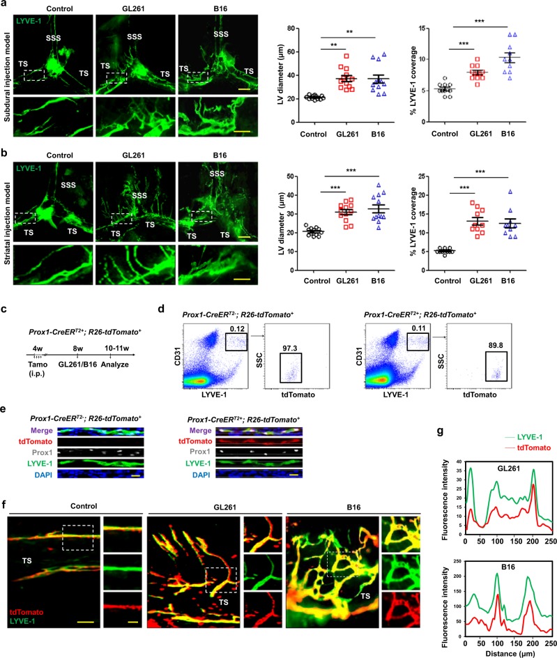
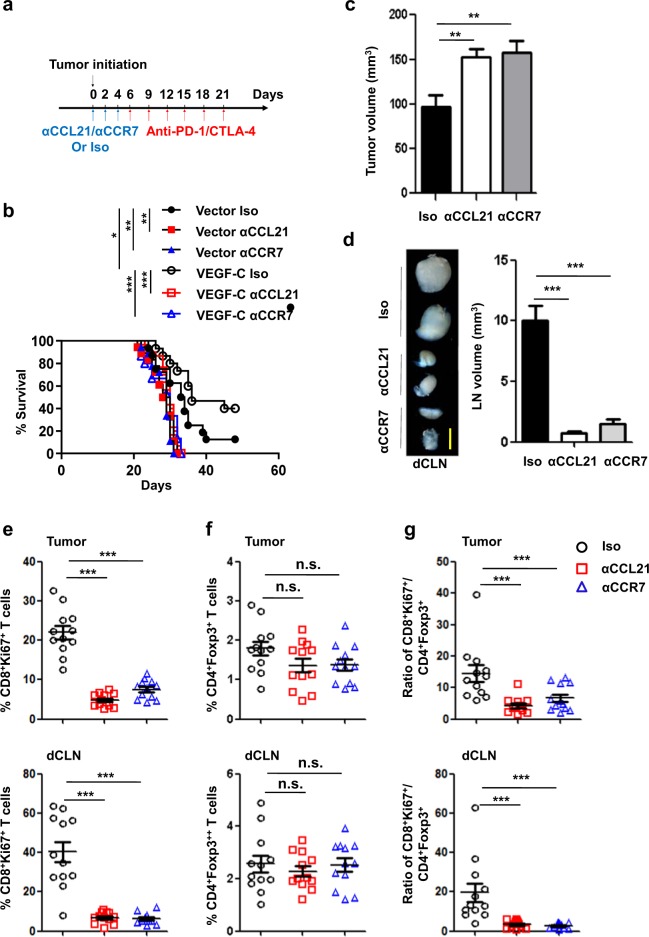
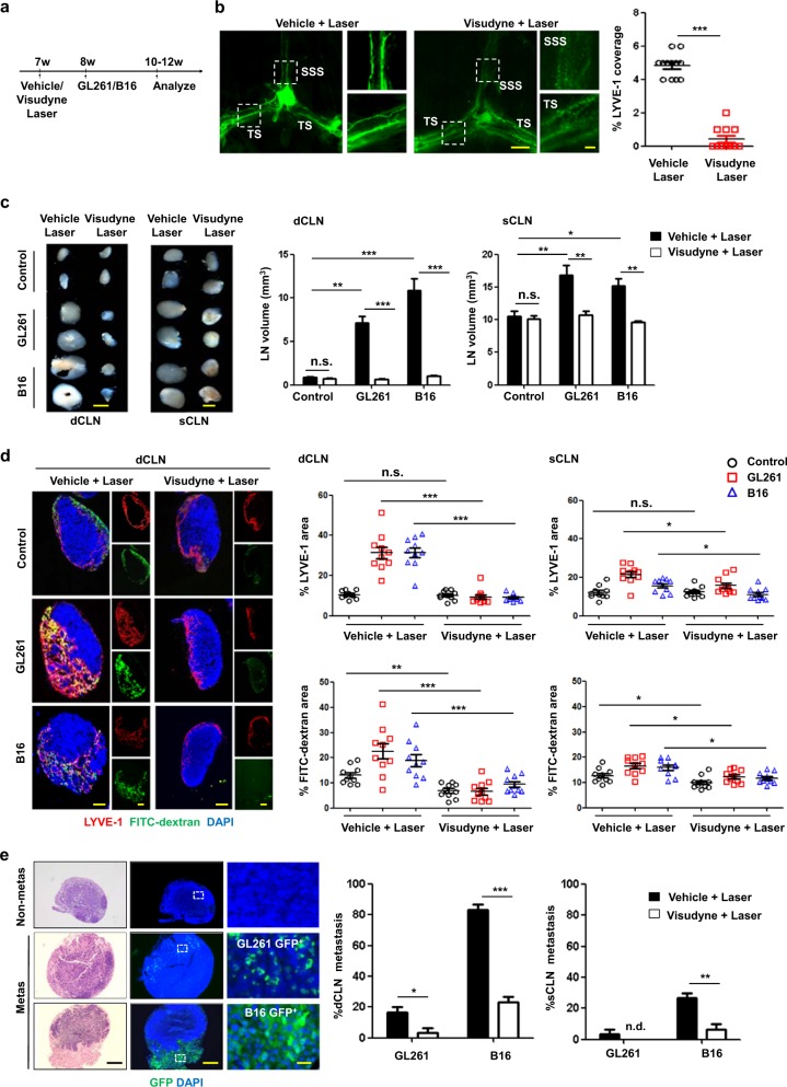
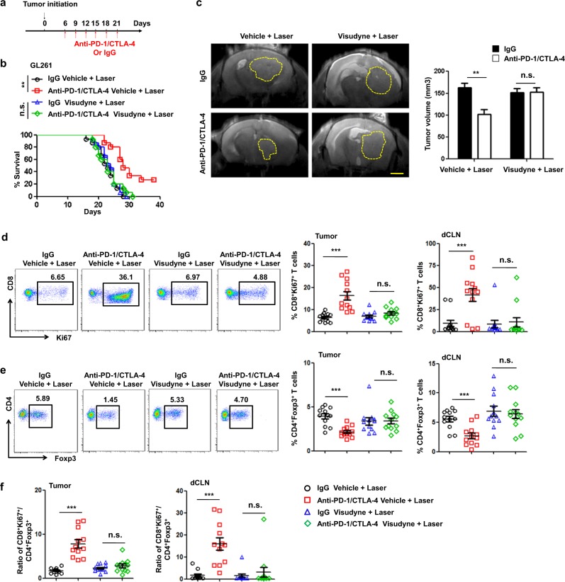
Recent studies have shown that meningeal lymphatic vessels (MLVs), which are located both dorsally and basally beneath the skull, provide a route for draining macromolecules and trafficking immune cells from the central nervous system (CNS) into cervical lymph nodes (CLNs), and thus represent a potential therapeutic target for treating neurodegenerative and neuroinflammatory diseases. However, the roles of MLVs in brain tumor drainage and immunity remain unexplored. Here we show that dorsal MLVs undergo extensive remodeling in mice with intracranial gliomas or metastatic melanomas. RNA-seq analysis of MLV endothelial cells revealed changes in the gene sets involved in lymphatic remodeling, fluid drainage, as well as inflammatory and immunological responses. Disruption of dorsal MLVs alone impaired intratumor fluid drainage and the dissemination of brain tumor cells to deep CLNs (dCLNs). Notably, the dendritic cell (DC) trafficking from intracranial tumor tissues to dCLNs decreased in mice with defective dorsal MLVs, and increased in mice with enhanced dorsal meningeal lymphangiogenesis. Strikingly, disruption of dorsal MLVs alone, without affecting basal MLVs or nasal LVs, significantly reduced the efficacy of combined anti-PD-1/CTLA-4 checkpoint therapy in striatal tumor models. Furthermore, mice bearing tumors overexpressing VEGF-C displayed a better response to anti-PD-1/CTLA-4 combination therapy, and this was abolished by CCL21/CCR7 blockade, suggesting that VEGF-C potentiates checkpoint therapy via the CCL21/CCR7 pathway. Together, the results of our study not only demonstrate the functional aspects of MLVs as classic lymphatic vasculature, but also highlight that they are essential in generating an efficient immune response against brain tumors.
最近的研究表明,脑膜淋巴管(MLVs)位于颅底和颅背,为大分子物质从中枢神经系统(CNS)引流到颈部淋巴结(CLNs)以及免疫细胞运输提供了途径,因此代表了治疗神经退行性和神经炎症性疾病的潜在治疗靶点。然而,MLVs 在脑肿瘤引流和免疫中的作用仍未被探索。在这里,我们发现在颅内神经胶质瘤或转移性黑色素瘤小鼠中,背侧 MLVs 经历了广泛的重塑。对 MLV 内皮细胞的 RNA-seq 分析显示,与淋巴管重塑、流体引流以及炎症和免疫反应相关的基因集发生了变化。单独破坏背侧 MLVs 会损害肿瘤内液体引流和脑肿瘤细胞向深部 CLNs(dCLNs)的扩散。值得注意的是,与具有正常背侧 MLVs 的小鼠相比,具有缺陷的背侧 MLVs 的小鼠颅内肿瘤组织中树突状细胞(DC)向 dCLNs 的迁移减少,而增强背侧脑膜淋巴管生成的小鼠则增加。引人注目的是,单独破坏背侧 MLVs,而不影响基底 MLVs 或鼻侧 LVs,会显著降低联合抗 PD-1/CTLA-4 检查点治疗在纹状体肿瘤模型中的疗效。此外,过表达 VEGF-C 的肿瘤小鼠对抗 PD-1/CTLA-4 联合治疗的反应更好,而阻断 CCL21/CCR7 则消除了这种反应,这表明 VEGF-C 通过 CCL21/CCR7 途径增强了检查点治疗。总之,我们的研究结果不仅证明了 MLVs 作为经典淋巴管的功能方面,而且还强调了它们在产生针对脑肿瘤的有效免疫反应方面的重要性。