Machida Takafumi, Machida Noboru
Department of Pharmacology, St. Marianna University School of Medicine, Kawasaki, Kanagawa 216-8511, Japan.
Laboratory of Veterinary Clinical Oncology, Tokyo University of Agriculture and Technology, Fuchu, Tokyo 183-8509, Japan.
Case Rep Vet Med. 2020 Feb 11;2020:5382687. doi: 10.1155/2020/5382687. eCollection 2020.
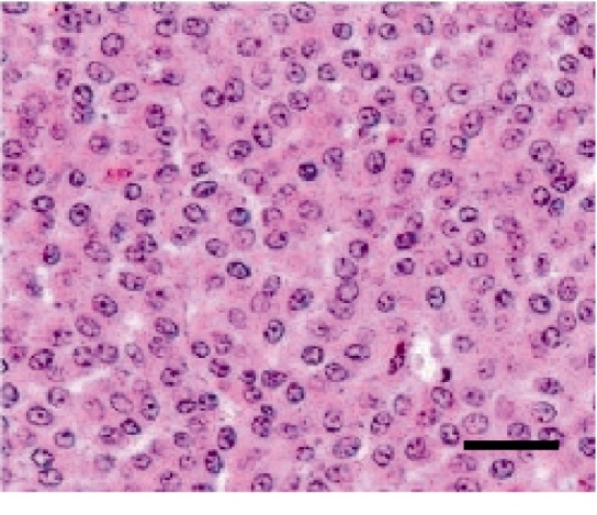
Pheochromocytomas are catecholamine-secreting tumors that are composed of neuroectoderm-derived chromaffin cells. An 8-year-old miniature dachshund with abdominal distension was diagnosed with a neuroendocrine tumor with invasion from the caudal vena cava to the right ventricular cavity. The dog died due to hypotensive shock from the vagal reflex, and on autopsy, an extra-adrenal pheochromocytoma (paraganglioma) was diagnosed in the caudal abdomen. At autopsy, the tumor plug of the caudal vena cava was confirmed. To the best of our knowledge, this is the first case report that echo-captured the extension of pheochromocytoma in the right ventricle and shows it in a figure and video file.
嗜铬细胞瘤是一种由神经外胚层来源的嗜铬细胞分泌儿茶酚胺的肿瘤。一只8岁患有腹胀的迷你腊肠犬被诊断患有神经内分泌肿瘤,肿瘤已从尾腔静脉侵犯至右心室腔。这只狗因迷走神经反射导致的低血压休克死亡,尸检时在腹部尾部诊断出一例肾上腺外嗜铬细胞瘤(副神经节瘤)。尸检时,证实了尾腔静脉有肿瘤栓子。据我们所知,这是第一例通过超声捕捉到嗜铬细胞瘤向右心室扩展并以图片和视频文件形式展示的病例报告。