Medical University of Vienna, Center for Medical Physics and Biomedical Engineering, Vienna, Austria.
Medical University of Vienna, Christian Doppler Laboratory OPTRAMED, Vienna, Austria.
J Biomed Opt. 2020 Feb;25(7):1-7. doi: 10.1117/1.JBO.25.7.071202.
5-Aminolevulinic acid (5-ALA)-based fluorescence guidance in conventional neurosurgical microscopes is limited to strongly fluorescent tumor tissue. Therefore, more sensitive, intrasurgical 5-ALA fluorescence visualization is needed.
Macroscopic fluorescence lifetime imaging (FLIM) was performed ex vivo on 5-ALA-labeled human glioma tissue through a surgical microscope to evaluate its feasibility and to compare it to fluorescence intensity imaging.
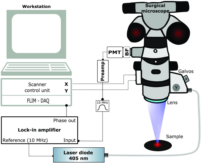
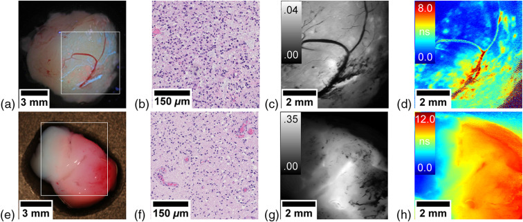
Frequency-domain FLIM was integrated into a surgical microscope, which enabled parallel wide-field white-light and fluorescence imaging. We first characterized our system and performed imaging of two samples of suspected low-grade glioma, which were compared to histopathology.
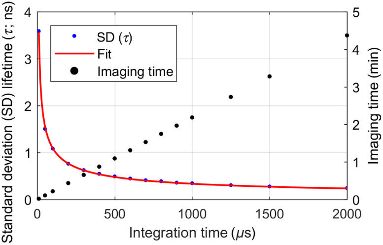
Our imaging system enabled macroscopic FLIM of a 6.5 × 6.5 mm2 field of view at spatial resolutions <20 μm. A frame of 512 × 512 pixels with a lifetime accuracy <1 ns was obtained in 65 s. Compared to conventional fluorescence imaging, FLIM considerably highlighted areas with weak 5-ALA fluorescence, which was in good agreement with histopathology.
Integration of macroscopic FLIM into a surgical microscope is feasible and a promising method for improved tumor delineation.
基于 5-氨基酮戊酸(5-ALA)的荧光引导在传统神经外科显微镜下仅限于强荧光肿瘤组织。因此,需要更敏感的、术中的 5-ALA 荧光可视化。
通过手术显微镜对 5-ALA 标记的人类脑胶质瘤组织进行离体宏观荧光寿命成像(FLIM),以评估其可行性,并将其与荧光强度成像进行比较。
频域 FLIM 被集成到手术显微镜中,该显微镜能够进行平行的宽场白光和荧光成像。我们首先对我们的系统进行了表征,并对两个疑似低级别胶质瘤样本进行了成像,然后将其与组织病理学进行了比较。
我们的成像系统能够对 6.5 × 6.5 毫米 2 的视场进行宏观 FLIM,空间分辨率<20 微米。在 65 秒内获得了具有<1 纳秒精度的 512 × 512 像素的一帧。与传统荧光成像相比,FLIM 相当大地突出了具有弱 5-ALA 荧光的区域,这与组织病理学非常吻合。
将宏观 FLIM 集成到手术显微镜中是可行的,并且是一种用于改善肿瘤描绘的有前途的方法。