Cao Xiao-Yan, Chen Cheng, Tian Na, Dong Xiang, Liang Xing, Xu Li-Jun, Cheng Cheng-Kung
School of Instrumentation and Optoelectronic Engineering, Beihang University, Beijing, 100191, China.
School of Biological Science and Medical Engineering, Beihang University, Beijing, 100191, China.
J Orthop Translat. 2020 Jan 14;21:81-90. doi: 10.1016/j.jot.2019.12.008. eCollection 2020 Mar.
Biodegradable suture anchors are commonly used for repairing torn rotator cuffs, but these biodegradable materials still suffer from low mechanical strength, poor osteointegration, and the generation of acidic degradation byproducts.
The purpose of this study was to evaluate the long-term mechanical behavior and osteogenetic capabilities of a biocomposite anchor injection molded with 30% β-tricalcium phosphate microparticles blended with 70% poly (L-lactide-co-glycolide) (85/15). This study investigated degradation and bone formation in a canine model. The initial mechanical behavior, mechanical strength retention with degradation time, and degradation features were investigated.
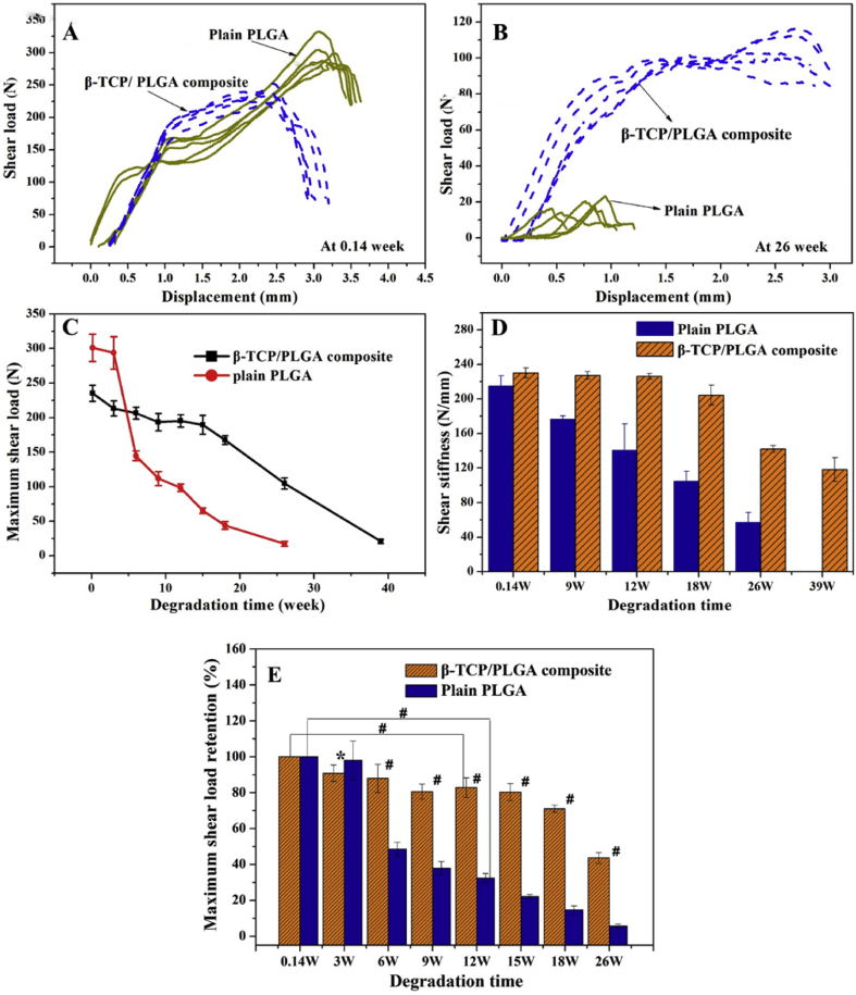
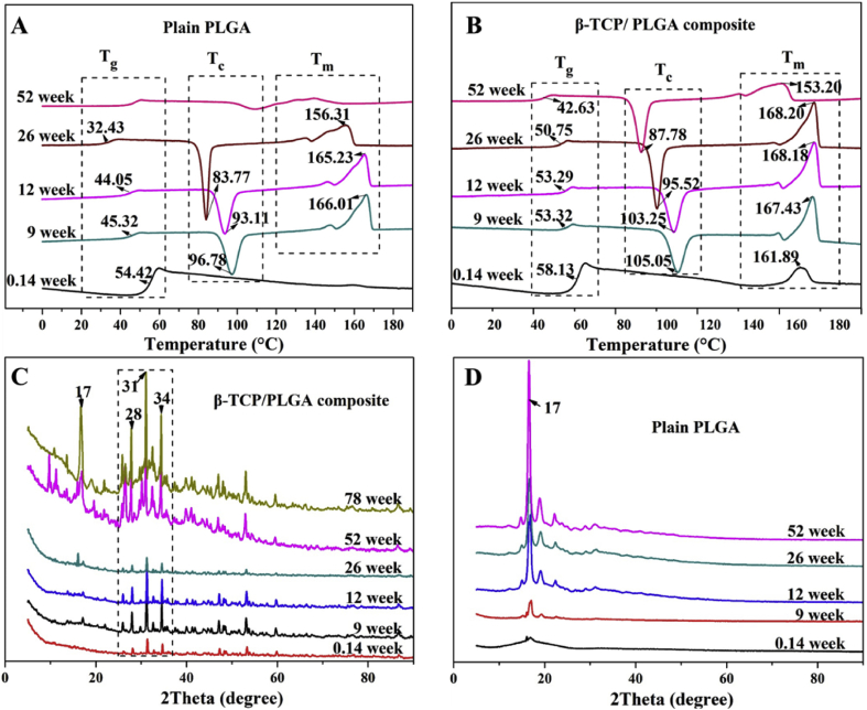
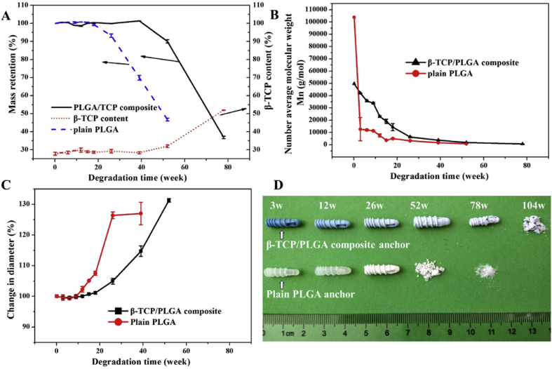
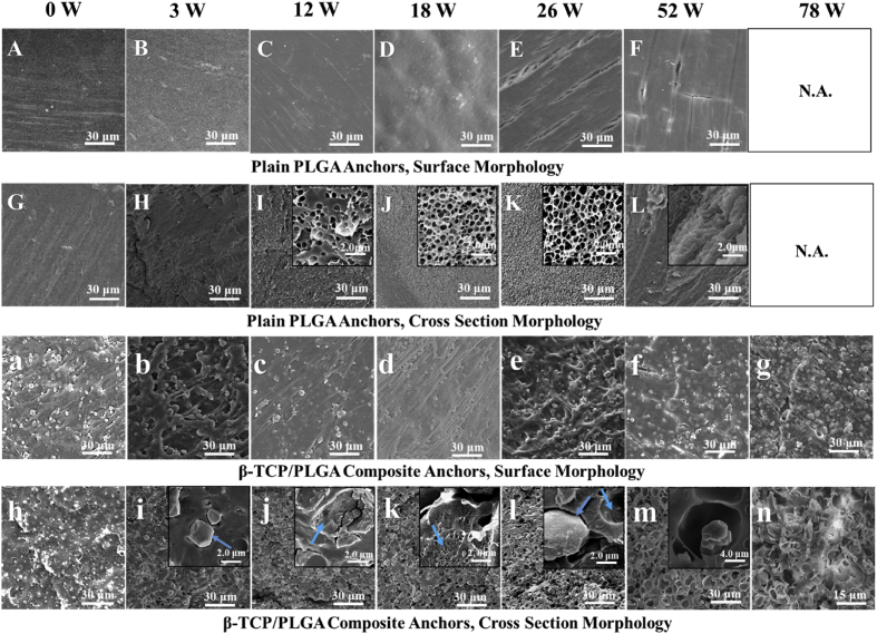
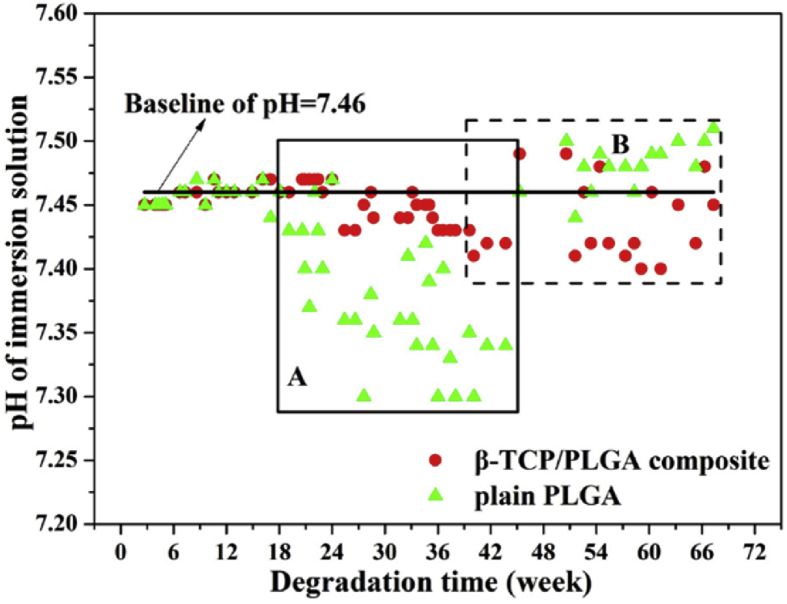
The results showed that the biocomposite anchor had sufficient initial mechanical stability confirmed by comparing the initial shear load on the anchor with the minimum shear load borne by an ankle fracture fixation screw, which is considered a worst-case implantation site for mechanical loading. The maximum shear load retention of the biocomposite anchor was 83% at 12 weeks, which is desirable, as it aligns with the rate of bone healing. The β-tricalcium phosphate fillers were evenly dispersed in the polymeric matrix and acted to slow the degradation rate and improve the mechanical strength of the anchor. The interface characteristics between the β-tricalcium phosphate particles and the polymeric matrix changed the degradation behavior of the biocomposite. Phosphate buffer saline was shown to diffuse through the interface into the biocomposite to inhibit the core accelerated degradation rate. , the addition of β-tricalcium phosphate induced new bone formation. The biocomposite material developed in this study demonstrated improved osteogenesis in comparison to a plain poly (L-lactide-co-glycolide) material. Neither anchor produced adverse tissue reactions, indicating that the biocomposite had favorable biocompatibility following long-term implantation.
In summary, the new biocomposite anchor presented in this study had favorable osteogenetic capability, mechanical property, and controlled degradation rate for bone fixation.
The new biocomposite anchor had sufficient initial and long-term fixation stability and bone formation capability in the canine model. It is indicated that the new biocomposite anchor has a potential for orthopedic application.
可生物降解的缝合锚钉常用于修复撕裂的肩袖,但这些可生物降解材料仍存在机械强度低、骨整合性差以及产生酸性降解副产物的问题。
本研究的目的是评估一种生物复合材料锚钉的长期力学行为和成骨能力,该锚钉通过注塑成型,由30%的β-磷酸三钙微粒与70%的聚(L-丙交酯-共-乙交酯)(85/15)混合而成。本研究在犬模型中研究了其降解和骨形成情况。研究了初始力学行为、力学强度随降解时间的保留情况以及降解特征。
结果表明,通过将锚钉上的初始剪切力与踝关节骨折固定螺钉承受的最小剪切力进行比较,证实了生物复合材料锚钉具有足够的初始力学稳定性,踝关节骨折固定螺钉承受的最小剪切力被认为是机械负荷的最坏植入部位。生物复合材料锚钉在12周时的最大剪切力保留率为83%,这是理想的,因为它与骨愈合速度一致。β-磷酸三钙填料均匀分散在聚合物基体中,起到减缓降解速度和提高锚钉机械强度的作用。β-磷酸三钙颗粒与聚合物基体之间的界面特性改变了生物复合材料的降解行为。磷酸盐缓冲盐水被证明可通过界面扩散到生物复合材料中,以抑制核心加速降解速度。此外,β-磷酸三钙的添加诱导了新骨形成。与普通聚(L-丙交酯-共-乙交酯)材料相比,本研究开发的生物复合材料表现出更好的成骨能力。两种锚钉均未产生不良组织反应,表明该生物复合材料在长期植入后具有良好的生物相容性。
总之,本研究中提出的新型生物复合材料锚钉在骨固定方面具有良好的成骨能力、力学性能和可控的降解速度。
新型生物复合材料锚钉在犬模型中具有足够的初始和长期固定稳定性以及骨形成能力。表明新型生物复合材料锚钉具有骨科应用潜力。