• 文献检索
  • 文档翻译
  • 深度研究
  • 学术资讯
  • Suppr Zotero 插件Zotero 插件
  • 邀请有礼
  • 套餐&价格
  • 历史记录
应用&插件
Suppr Zotero 插件Zotero 插件浏览器插件Mac 客户端Windows 客户端微信小程序
定价
高级版会员购买积分包购买API积分包
服务
文献检索文档翻译深度研究API 文档MCP 服务
关于我们
关于 Suppr公司介绍联系我们用户协议隐私条款
关注我们

Suppr 超能文献

核心技术专利:CN118964589B侵权必究
粤ICP备2023148730 号-1Suppr @ 2026

文献检索

告别复杂PubMed语法,用中文像聊天一样搜索,搜遍4000万医学文献。AI智能推荐,让科研检索更轻松。

立即免费搜索

文件翻译

保留排版,准确专业,支持PDF/Word/PPT等文件格式,支持 12+语言互译。

免费翻译文档

深度研究

AI帮你快速写综述,25分钟生成高质量综述,智能提取关键信息,辅助科研写作。

立即免费体验

P2X7 C 端区的进化起源:在远古有颌脊椎动物中,一个类似 P2X4 的基因捕获了一个古老的压舱物结构域。

Evolutionary Origin of the P2X7 C-ter Region: Capture of an Ancient Ballast Domain by a P2X4-Like Gene in Ancient Jawed Vertebrates.

机构信息

Immunology Unit, Department of Chemistry and Biotechnology, Tallinn University of Technology, Tallinn, Estonia.

Department of Chemistry and Biotechnology, Tallinn University of Technology, Tallinn, Estonia.

出版信息

Front Immunol. 2020 Feb 6;11:113. doi: 10.3389/fimmu.2020.00113. eCollection 2020.

DOI:10.3389/fimmu.2020.00113
PMID:32117264
原文链接:https://pmc.ncbi.nlm.nih.gov/articles/PMC7016195/
Abstract

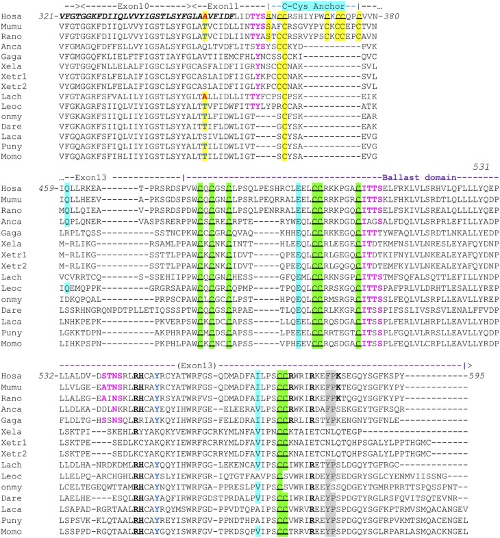
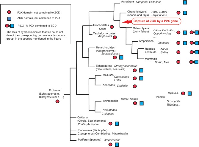
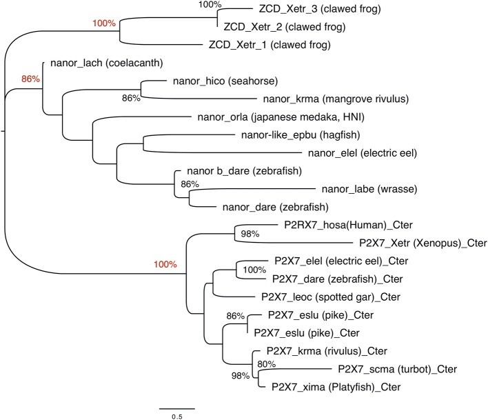
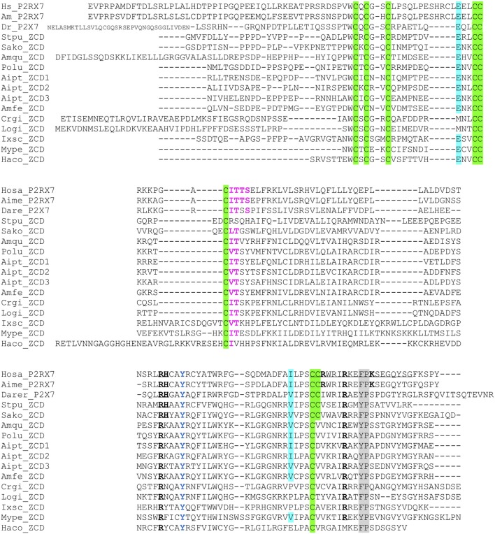
P2X purinergic receptors are extracellular ATP-gated ion channel receptors present on the cell plasma membrane. P2X receptors have been found in Metazoa, fungi, amoebas, and in plants. In mammals, P2X7 is expressed by a large number of cell types and is involved in inflammation and immunity. Remarkably, P2X7 does not desensitize as other P2X do, a feature linked to a "C-cysteine anchor" intra-cytoplasmic motif encoded by exon 11. Another specific feature of P2X7 is its C-terminal cytoplasmic ballast domain (exon 13) which contains a zinc (Zn) coordinating cysteine motif and a GDP-binding region. To determine the origin of P2X7, we analyzed and compared sequences and protein motifs of the C-terminal intra-cytoplasmic region across all main groups of Metazoa. We identified proteins with typical ballast domains, sharing a remarkably conserved Zn-coordinating cysteine motif. Apart from vertebrates, these ballast domains were not associated with a typical P2X architecture. These results strongly suggest that P2X7 resulted from the fusion of a P2X gene, highly similar to P2X4, with an exon encoding a ballast domain. Our work brings new evidence on the origin of the P2X7 purinergic receptor and identifies the Zn-coordinating cysteine domain as the fundamental feature of the ancient ballast fold.

摘要

P2X 嘌呤能受体是位于细胞质膜上的细胞外 ATP 门控离子通道受体。P2X 受体已在后生动物、真菌、变形虫和植物中发现。在哺乳动物中,大量细胞类型表达 P2X7,参与炎症和免疫。值得注意的是,P2X7 不像其他 P2X 那样脱敏,这一特征与由外显子 11 编码的“C-半胱氨酸锚”细胞内基序有关。P2X7 的另一个特有特征是其 C 端细胞质压舱物域(外显子 13),其中包含一个锌(Zn)配位半胱氨酸基序和一个 GDP 结合区域。为了确定 P2X7 的起源,我们分析并比较了后生动物所有主要类群中跨膜内 C 末端区域的序列和蛋白基序。我们确定了具有典型压舱物域的蛋白,这些蛋白共享一个非常保守的 Zn 配位半胱氨酸基序。除了脊椎动物外,这些压舱物域与典型的 P2X 结构没有关联。这些结果强烈表明 P2X7 是由与 P2X4 高度相似的 P2X 基因与编码压舱物域的外显子融合产生的。我们的工作为 P2X7 嘌呤能受体的起源提供了新的证据,并将 Zn 配位半胱氨酸基序确定为古老压舱物折叠的基本特征。

https://cdn.ncbi.nlm.nih.gov/pmc/blobs/d254/7016195/b07dcdfbe5cd/fimmu-11-00113-g0008.jpg
https://cdn.ncbi.nlm.nih.gov/pmc/blobs/d254/7016195/d57a4c2c591c/fimmu-11-00113-g0001.jpg
https://cdn.ncbi.nlm.nih.gov/pmc/blobs/d254/7016195/9344397aff57/fimmu-11-00113-g0002.jpg
https://cdn.ncbi.nlm.nih.gov/pmc/blobs/d254/7016195/e9b95936ddf7/fimmu-11-00113-g0003.jpg
https://cdn.ncbi.nlm.nih.gov/pmc/blobs/d254/7016195/0ef268624f00/fimmu-11-00113-g0004.jpg
https://cdn.ncbi.nlm.nih.gov/pmc/blobs/d254/7016195/a4ce613cca65/fimmu-11-00113-g0005.jpg
https://cdn.ncbi.nlm.nih.gov/pmc/blobs/d254/7016195/41c8f609cd08/fimmu-11-00113-g0006.jpg
https://cdn.ncbi.nlm.nih.gov/pmc/blobs/d254/7016195/6f3be2f01708/fimmu-11-00113-g0007.jpg
https://cdn.ncbi.nlm.nih.gov/pmc/blobs/d254/7016195/b07dcdfbe5cd/fimmu-11-00113-g0008.jpg

相似文献

1
Evolutionary Origin of the P2X7 C-ter Region: Capture of an Ancient Ballast Domain by a P2X4-Like Gene in Ancient Jawed Vertebrates.P2X7 C 端区的进化起源:在远古有颌脊椎动物中,一个类似 P2X4 的基因捕获了一个古老的压舱物结构域。
Front Immunol. 2020 Feb 6;11:113. doi: 10.3389/fimmu.2020.00113. eCollection 2020.
2
How Structural Biology Has Directly Impacted Our Understanding of P2X Receptor Function and Gating.结构生物学如何直接影响我们对 P2X 受体功能和门控的理解。
Methods Mol Biol. 2022;2510:1-29. doi: 10.1007/978-1-0716-2384-8_1.
3
New insights into P2X7 receptor regulation: Ca-calmodulin and GDP bind to the soluble P2X7 ballast domain.对 P2X7 受体调节的新认识:Ca-钙调蛋白和 GDP 结合到可溶性 P2X7 压舱域。
J Biol Chem. 2022 Oct;298(10):102495. doi: 10.1016/j.jbc.2022.102495. Epub 2022 Sep 15.
4
Ionotropic Purinergic Receptors P2X4 and P2X7: Proviral or Antiviral? An Insight into P2X Receptor Signaling and Hepatitis C Virus Infection.离子型嘌呤能受体P2X4和P2X7:促进病毒还是抗病毒?深入了解P2X受体信号传导与丙型肝炎病毒感染
Viral Immunol. 2016 Sep;29(7):401-8. doi: 10.1089/vim.2016.0008. Epub 2016 Aug 15.
5
Functional expression of P2X1, P2X4 and P2X7 purinergic receptors in human monocyte-derived macrophages.人源单核细胞衍生巨噬细胞中 P2X1、P2X4 和 P2X7 嘌呤能受体的功能表达。
Eur J Pharmacol. 2020 Dec 5;888:173460. doi: 10.1016/j.ejphar.2020.173460. Epub 2020 Aug 14.
6
Structural and Functional Features of the P2X4 Receptor: An Immunological Perspective.P2X4 受体的结构和功能特征:免疫学视角。
Front Immunol. 2021 Mar 25;12:645834. doi: 10.3389/fimmu.2021.645834. eCollection 2021.
7
Lithocholic acid inhibits P2X2 and potentiates P2X4 receptor channel gating.胆酸抑制 P2X2 并增强 P2X4 受体通道门控。
J Steroid Biochem Mol Biol. 2020 Sep;202:105725. doi: 10.1016/j.jsbmb.2020.105725. Epub 2020 Jul 8.
8
P2X purinergic receptor channel expression and function in bovine aortic endothelium.P2X嘌呤能受体通道在牛主动脉内皮中的表达与功能
Am J Physiol Heart Circ Physiol. 2002 Jun;282(6):H2106-16. doi: 10.1152/ajpheart.00892.2001.
9
A dual polybasic motif determines phosphoinositide binding and regulation in the P2X channel family.双碱性基序决定 P2X 通道家族中磷酯酰肌醇的结合和调节。
PLoS One. 2012;7(7):e40595. doi: 10.1371/journal.pone.0040595. Epub 2012 Jul 11.
10
Deviant reporter expression and P2X4 passenger gene overexpression in the soluble EGFP BAC transgenic P2X7 reporter mouse model.畸形 reporter 表达和 P2X4 乘客基因过表达在可溶性 EGFP BAC 转基因 P2X7 reporter 小鼠模型中。
Sci Rep. 2020 Nov 16;10(1):19876. doi: 10.1038/s41598-020-76428-0.

引用本文的文献

1
P2X7 regulates ependymo-radial glial cell proliferation in adult Danio rerio following spinal cord injury.P2X7 调控成年斑马鱼脊髓损伤后室管膜-放射状胶质细胞的增殖。
Biol Open. 2024 Apr 15;13(4). doi: 10.1242/bio.060270. Epub 2024 Apr 11.
2
Tumour immune escape via P2X7 receptor signalling.肿瘤通过 P2X7 受体信号逃逸免疫。
Front Immunol. 2023 Oct 30;14:1287310. doi: 10.3389/fimmu.2023.1287310. eCollection 2023.
3
Epithelial wound healing in Clytia hemisphaerica provides insights into extracellular ATP signaling mechanisms and P2XR evolution.

本文引用的文献

1
Full-Length P2X Structures Reveal How Palmitoylation Prevents Channel Desensitization.全长 P2X 结构揭示了棕榈酰化如何防止通道脱敏。
Cell. 2019 Oct 17;179(3):659-670.e13. doi: 10.1016/j.cell.2019.09.017. Epub 2019 Oct 3.
2
P2X7 Interactions and Signaling - Making Head or Tail of It.P2X7相互作用与信号传导——弄清楚其中的头绪
Front Mol Neurosci. 2019 Aug 7;12:183. doi: 10.3389/fnmol.2019.00183. eCollection 2019.
3
The P2X7 Receptor in Inflammatory Diseases: Angel or Demon?炎症性疾病中的P2X7受体:天使还是恶魔?
克利特海葵的上皮细胞伤口愈合为细胞外 ATP 信号机制和 P2XR 进化提供了新见解。
Sci Rep. 2023 Nov 1;13(1):18819. doi: 10.1038/s41598-023-45424-5.
4
Animal Models for the Investigation of P2X7 Receptors.用于研究 P2X7 受体的动物模型。
Int J Mol Sci. 2023 May 4;24(9):8225. doi: 10.3390/ijms24098225.
5
A unique eukaryotic lineage of composite-like DNA transposons encoding a DDD/E transposase and a His-Me finger homing endonuclease.一种独特的真核生物复合样DNA转座子谱系,其编码一种DDD/E转座酶和一种组氨酸-甲硫氨酸指归巢内切核酸酶。
Mob DNA. 2022 Oct 22;13(1):24. doi: 10.1186/s13100-022-00281-3.
6
Corrigendum: Purinergic signaling systems across comparative models of spinal cord injury.勘误:跨脊髓损伤比较模型的嘌呤能信号系统。
Neural Regen Res. 2023 Mar;18(3):689-696. doi: 10.4103/1673-5374.350234.
7
The P2X7R-NLRP3 and AIM2 Inflammasome Platforms Mark the Complexity/Severity of Viral or Metabolic Liver Damage.P2X7R-NLRP3 和 AIM2 炎性小体平台标志着病毒或代谢性肝损伤的复杂性/严重程度。
Int J Mol Sci. 2022 Jul 4;23(13):7447. doi: 10.3390/ijms23137447.
8
Purinergic signaling systems across comparative models of spinal cord injury.跨脊髓损伤比较模型的嘌呤能信号系统
Neural Regen Res. 2022 Nov;17(11):2391-2398. doi: 10.4103/1673-5374.338993.
9
P2X7 Variants in Oncogenesis.P2X7变体在肿瘤发生中的作用
Cells. 2021 Jan 19;10(1):189. doi: 10.3390/cells10010189.
10
P2X7 Receptor Upregulation in Huntington's Disease Brains.亨廷顿病大脑中P2X7受体上调
Front Mol Neurosci. 2020 Oct 6;13:567430. doi: 10.3389/fnmol.2020.567430. eCollection 2020.
Front Pharmacol. 2018 Feb 6;9:52. doi: 10.3389/fphar.2018.00052. eCollection 2018.
4
The Elusive P2X7 Macropore. elusive P2X7 巨孔
Trends Cell Biol. 2018 May;28(5):392-404. doi: 10.1016/j.tcb.2018.01.005. Epub 2018 Feb 10.
5
P2X4: A fast and sensitive purinergic receptor.P2X4:一种快速而敏感的嘌呤能受体。
Biomed J. 2017 Oct;40(5):245-256. doi: 10.1016/j.bj.2017.06.010. Epub 2017 Nov 10.
6
The P2X7 receptor forms a dye-permeable pore independent of its intracellular domain but dependent on membrane lipid composition.P2X7 受体形成一种染料渗透性孔道,该孔道不依赖其细胞内结构域,但依赖于膜脂组成。
Elife. 2017 Sep 18;6:e31186. doi: 10.7554/eLife.31186.
7
The P2X7 Receptor in Infection and Inflammation.P2X7 受体在感染和炎症中的作用。
Immunity. 2017 Jul 18;47(1):15-31. doi: 10.1016/j.immuni.2017.06.020.
8
Structural basis for subtype-specific inhibition of the P2X7 receptor.P2X7受体亚型特异性抑制的结构基础。
Elife. 2016 Dec 9;5:e22153. doi: 10.7554/eLife.22153.
9
MEGA7: Molecular Evolutionary Genetics Analysis Version 7.0 for Bigger Datasets.MEGA7:适用于更大数据集的分子进化遗传学分析版本7.0
Mol Biol Evol. 2016 Jul;33(7):1870-4. doi: 10.1093/molbev/msw054. Epub 2016 Mar 22.
10
Comparative study of the P2X gene family in animals and plants.动物和植物中P2X基因家族的比较研究。
Purinergic Signal. 2016 Jun;12(2):269-81. doi: 10.1007/s11302-016-9501-z. Epub 2016 Feb 13.