• 文献检索
  • 文档翻译
  • 深度研究
  • 学术资讯
  • Suppr Zotero 插件Zotero 插件
  • 邀请有礼
  • 套餐&价格
  • 历史记录
应用&插件
Suppr Zotero 插件Zotero 插件浏览器插件Mac 客户端Windows 客户端微信小程序
定价
高级版会员购买积分包购买API积分包
服务
文献检索文档翻译深度研究API 文档MCP 服务
关于我们
关于 Suppr公司介绍联系我们用户协议隐私条款
关注我们

Suppr 超能文献

核心技术专利:CN118964589B侵权必究
粤ICP备2023148730 号-1Suppr @ 2026

文献检索

告别复杂PubMed语法,用中文像聊天一样搜索,搜遍4000万医学文献。AI智能推荐,让科研检索更轻松。

立即免费搜索

文件翻译

保留排版,准确专业,支持PDF/Word/PPT等文件格式,支持 12+语言互译。

免费翻译文档

深度研究

AI帮你快速写综述,25分钟生成高质量综述,智能提取关键信息,辅助科研写作。

立即免费体验

联合人工智能和放射科医生评估解读筛查性乳房 X 光照片的效果。

Evaluation of Combined Artificial Intelligence and Radiologist Assessment to Interpret Screening Mammograms.

机构信息

Computational Oncology, Sage Bionetworks, Seattle, Washington.

Kaiser Permanente Washington Health Research Institute, Seattle, Washington.

出版信息

JAMA Netw Open. 2020 Mar 2;3(3):e200265. doi: 10.1001/jamanetworkopen.2020.0265.

DOI:10.1001/jamanetworkopen.2020.0265
PMID:32119094
原文链接:https://pmc.ncbi.nlm.nih.gov/articles/PMC7052735/
Abstract

IMPORTANCE

Mammography screening currently relies on subjective human interpretation. Artificial intelligence (AI) advances could be used to increase mammography screening accuracy by reducing missed cancers and false positives.

OBJECTIVE

To evaluate whether AI can overcome human mammography interpretation limitations with a rigorous, unbiased evaluation of machine learning algorithms.

DESIGN, SETTING, AND PARTICIPANTS: In this diagnostic accuracy study conducted between September 2016 and November 2017, an international, crowdsourced challenge was hosted to foster AI algorithm development focused on interpreting screening mammography. More than 1100 participants comprising 126 teams from 44 countries participated. Analysis began November 18, 2016.

MAIN OUTCOMES AND MEASUREMENTS

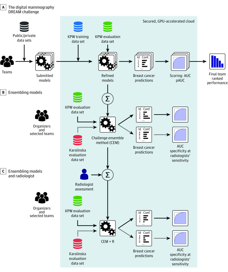
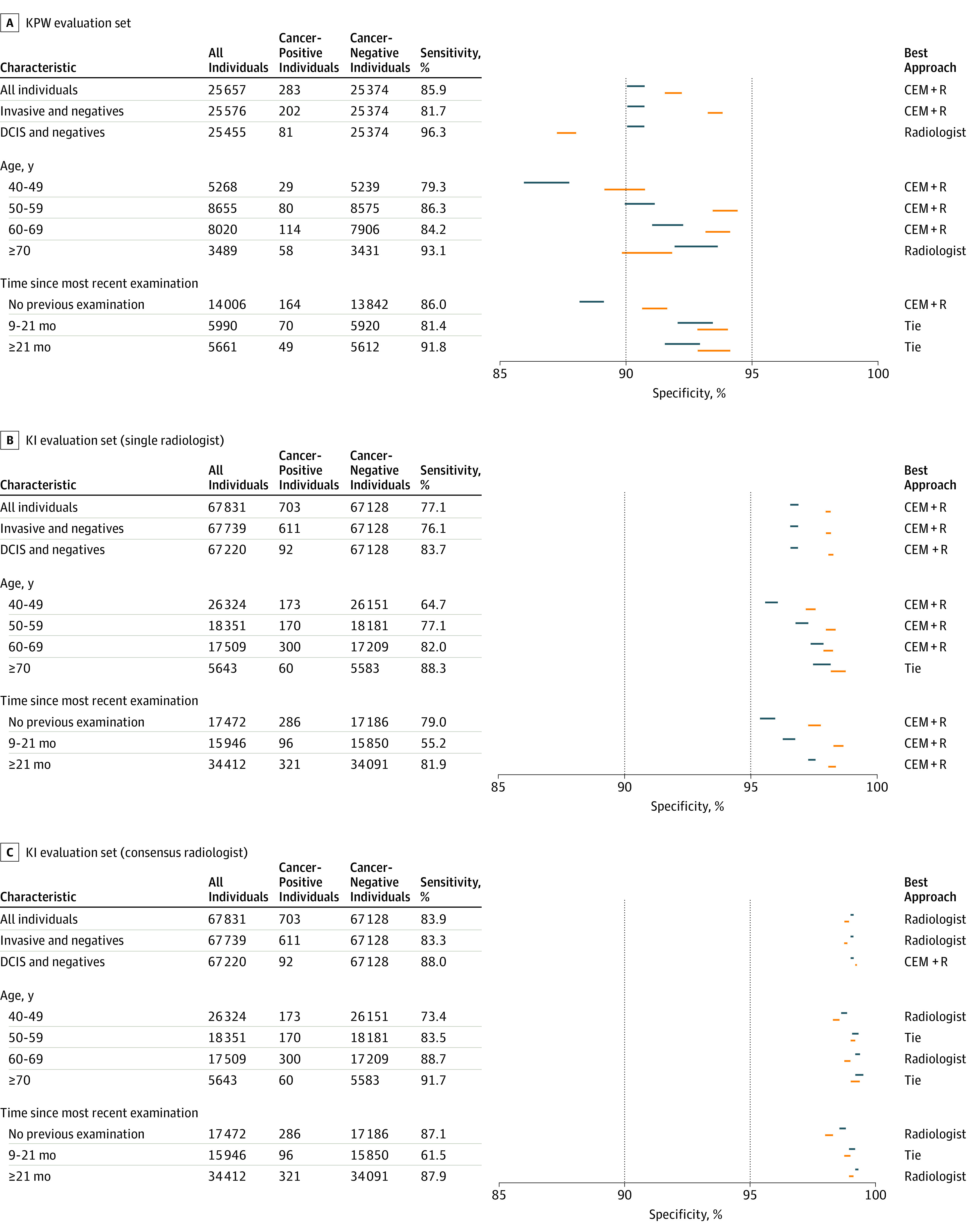
Algorithms used images alone (challenge 1) or combined images, previous examinations (if available), and clinical and demographic risk factor data (challenge 2) and output a score that translated to cancer yes/no within 12 months. Algorithm accuracy for breast cancer detection was evaluated using area under the curve and algorithm specificity compared with radiologists' specificity with radiologists' sensitivity set at 85.9% (United States) and 83.9% (Sweden). An ensemble method aggregating top-performing AI algorithms and radiologists' recall assessment was developed and evaluated.

RESULTS

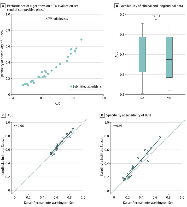
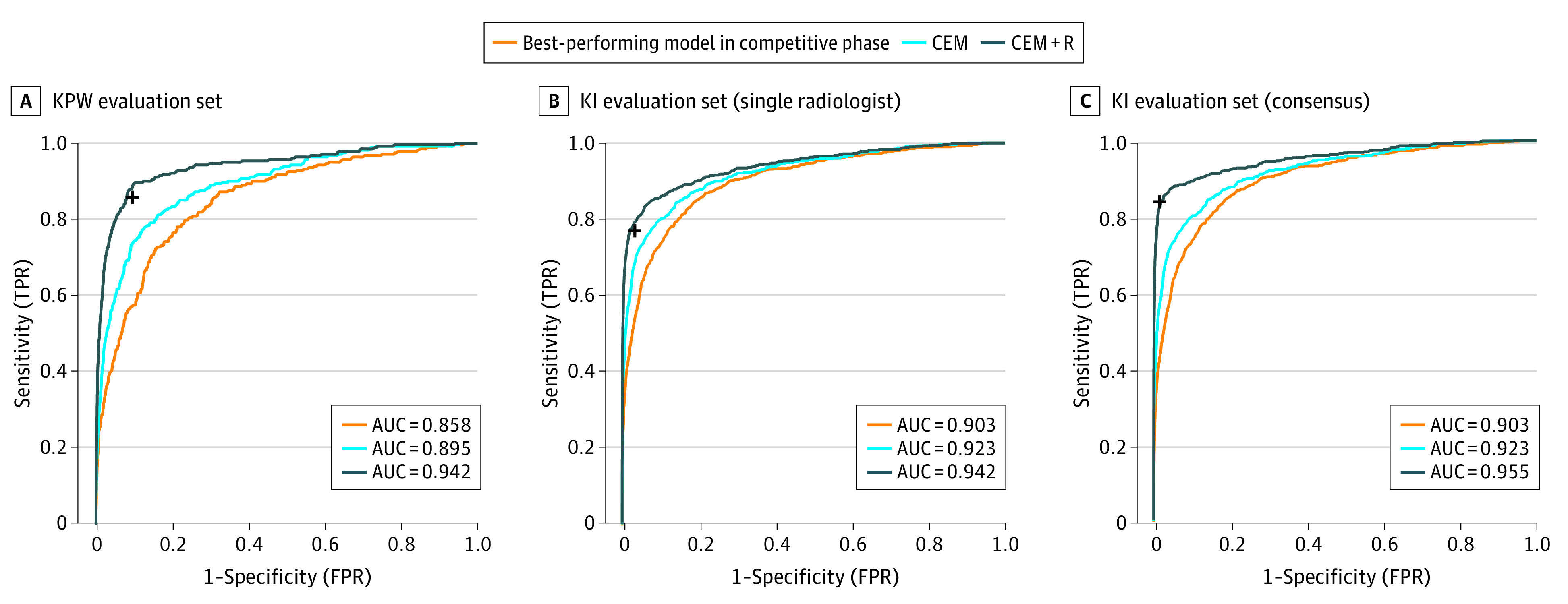
Overall, 144 231 screening mammograms from 85 580 US women (952 cancer positive ≤12 months from screening) were used for algorithm training and validation. A second independent validation cohort included 166 578 examinations from 68 008 Swedish women (780 cancer positive). The top-performing algorithm achieved an area under the curve of 0.858 (United States) and 0.903 (Sweden) and 66.2% (United States) and 81.2% (Sweden) specificity at the radiologists' sensitivity, lower than community-practice radiologists' specificity of 90.5% (United States) and 98.5% (Sweden). Combining top-performing algorithms and US radiologist assessments resulted in a higher area under the curve of 0.942 and achieved a significantly improved specificity (92.0%) at the same sensitivity.

CONCLUSIONS AND RELEVANCE

While no single AI algorithm outperformed radiologists, an ensemble of AI algorithms combined with radiologist assessment in a single-reader screening environment improved overall accuracy. This study underscores the potential of using machine learning methods for enhancing mammography screening interpretation.

摘要

重要性

乳腺 X 线摄影筛查目前依赖于主观的人工解释。人工智能(AI)的进步可以通过减少漏诊的癌症和假阳性来提高乳腺 X 线摄影筛查的准确性。

目的

通过对机器学习算法进行严格的、无偏的评估,评估 AI 是否可以克服人类乳腺 X 线摄影解释的局限性。

设计、环境和参与者: 在这项诊断准确性研究中,于 2016 年 9 月至 2017 年 11 月期间进行,举办了一项国际众包挑战赛,以促进专注于解释筛查性乳腺 X 线摄影的 AI 算法的开发。来自 44 个国家的 126 个团队的 1100 多名参与者参加了这项研究。分析于 2016 年 11 月 18 日开始。

主要结果和测量

算法仅使用图像(挑战 1)或结合图像、以前的检查(如果有)以及临床和人口统计学风险因素数据(挑战 2),并输出在 12 个月内癌症呈阳性/阴性的评分。使用曲线下面积评估算法检测乳腺癌的准确性,并与放射科医生的特异性进行比较,将放射科医生的敏感性设定为 85.9%(美国)和 83.9%(瑞典)。开发并评估了一种聚合表现最佳的 AI 算法和放射科医生的召回评估的集成方法。

结果

总体而言,使用了来自 85580 名美国女性的 144231 份筛查性乳腺 X 线摄影图像(12 个月内≤12 个月有 952 例癌症阳性)进行算法训练和验证。第二个独立验证队列包括来自 68008 名瑞典女性的 166578 次检查(780 例癌症阳性)。表现最佳的算法在美国的曲线下面积为 0.858,在瑞典为 0.903,在放射科医生设定的敏感性为 66.2%,在瑞典为 81.2%,特异性低于社区实践放射科医生的特异性 90.5%(美国)和 98.5%(瑞典)。将表现最佳的算法组合并结合美国放射科医生的评估,曲线下面积提高到 0.942,特异性(92.0%)在相同的敏感性下得到显著提高。

结论和相关性

虽然没有单一的 AI 算法超过放射科医生,但在单读筛查环境中,AI 算法的集合与放射科医生的评估相结合,提高了整体准确性。这项研究强调了使用机器学习方法增强乳腺 X 线摄影筛查解释的潜力。

https://cdn.ncbi.nlm.nih.gov/pmc/blobs/bfb3/7052735/ac91994dd16b/jamanetwopen-e200265-g004.jpg
https://cdn.ncbi.nlm.nih.gov/pmc/blobs/bfb3/7052735/50f42ff38208/jamanetwopen-e200265-g001.jpg
https://cdn.ncbi.nlm.nih.gov/pmc/blobs/bfb3/7052735/612692985476/jamanetwopen-e200265-g002.jpg
https://cdn.ncbi.nlm.nih.gov/pmc/blobs/bfb3/7052735/d9ba5ec55bdf/jamanetwopen-e200265-g003.jpg
https://cdn.ncbi.nlm.nih.gov/pmc/blobs/bfb3/7052735/ac91994dd16b/jamanetwopen-e200265-g004.jpg

相似文献

1
Evaluation of Combined Artificial Intelligence and Radiologist Assessment to Interpret Screening Mammograms.联合人工智能和放射科医生评估解读筛查性乳房 X 光照片的效果。
JAMA Netw Open. 2020 Mar 2;3(3):e200265. doi: 10.1001/jamanetworkopen.2020.0265.
2
External Evaluation of 3 Commercial Artificial Intelligence Algorithms for Independent Assessment of Screening Mammograms.商业人工智能算法独立评估筛查性乳房 X 光照片的外部评估。
JAMA Oncol. 2020 Oct 1;6(10):1581-1588. doi: 10.1001/jamaoncol.2020.3321.
3
Changes in cancer detection and false-positive recall in mammography using artificial intelligence: a retrospective, multireader study.人工智能在乳腺癌检测和假阳性召回中的变化:一项回顾性、多读者研究。
Lancet Digit Health. 2020 Mar;2(3):e138-e148. doi: 10.1016/S2589-7500(20)30003-0. Epub 2020 Feb 6.
4
Impact of AI for Digital Breast Tomosynthesis on Breast Cancer Detection and Interpretation Time.人工智能对数字乳腺断层合成术在乳腺癌检测和解读时间方面的影响。
Radiol Artif Intell. 2024 May;6(3):e230318. doi: 10.1148/ryai.230318.
5
Screening mammography performance according to breast density: a comparison between radiologists versus standalone intelligence detection.根据乳腺密度评估乳腺钼靶筛查性能:放射科医生与独立智能检测的比较。
Breast Cancer Res. 2024 Apr 22;26(1):68. doi: 10.1186/s13058-024-01821-w.
6
Improved Cancer Detection Using Artificial Intelligence: a Retrospective Evaluation of Missed Cancers on Mammography.利用人工智能提高癌症检测率:对乳腺 X 光摄影术漏诊癌症的回顾性评估。
J Digit Imaging. 2019 Aug;32(4):625-637. doi: 10.1007/s10278-019-00192-5.
7
External Validation of an Ensemble Model for Automated Mammography Interpretation by Artificial Intelligence.人工智能辅助自动化乳腺 X 线摄影判读的集成模型的外部验证。
JAMA Netw Open. 2022 Nov 1;5(11):e2242343. doi: 10.1001/jamanetworkopen.2022.42343.
8
Performance of artificial intelligence in 7533 consecutive prevalent screening mammograms from the BreastScreen Australia program.澳大利亚乳腺筛查计划中 7533 例连续现患筛查乳腺 X 线摄影的人工智能性能。
Eur Radiol. 2024 Jun;34(6):3947-3957. doi: 10.1007/s00330-023-10396-7. Epub 2023 Nov 13.
9
Effect of artificial intelligence-based computer-aided diagnosis on the screening outcomes of digital mammography: a matched cohort study.基于人工智能的计算机辅助诊断对数字乳腺 X 线摄影筛查结果的影响:一项匹配队列研究。
Eur Radiol. 2023 Oct;33(10):7186-7198. doi: 10.1007/s00330-023-09692-z. Epub 2023 May 16.
10
Stand-Alone Use of Artificial Intelligence for Digital Mammography and Digital Breast Tomosynthesis Screening: A Retrospective Evaluation.人工智能在数字乳腺 X 线摄影和数字乳腺断层合成筛查中的独立应用:一项回顾性评估。
Radiology. 2022 Mar;302(3):535-542. doi: 10.1148/radiol.211590. Epub 2021 Dec 14.

引用本文的文献

1
Interpreting Venous and Arterial Ulcer Images Through the Grad-CAM Lens: Insights and Implications in CNN-Based Wound Image Classification.通过Grad-CAM视角解读静脉和动脉溃疡图像:基于卷积神经网络的伤口图像分类中的见解与启示
Diagnostics (Basel). 2025 Aug 28;15(17):2184. doi: 10.3390/diagnostics15172184.
2
Combined use of two artificial intelligence-based algorithms for mammography triaging: a retrospective simulation study.两种基于人工智能的算法联合用于乳腺钼靶筛查分诊:一项回顾性模拟研究。
Eur Radiol. 2025 Aug 21. doi: 10.1007/s00330-025-11849-x.
3
Genotype Distribution and Migration Patterns of Hepatitis C Virus in Shandong Province, China: Molecular Epidemiology and Phylogenetic Study.

本文引用的文献

1
Improving Accuracy and Efficiency with Concurrent Use of Artificial Intelligence for Digital Breast Tomosynthesis.同时使用人工智能提高数字乳腺断层合成的准确性和效率。
Radiol Artif Intell. 2019 Jul 31;1(4):e180096. doi: 10.1148/ryai.2019180096.
2
Deep Neural Networks Improve Radiologists' Performance in Breast Cancer Screening.深度神经网络可提高放射科医生在乳腺癌筛查中的表现。
IEEE Trans Med Imaging. 2020 Apr;39(4):1184-1194. doi: 10.1109/TMI.2019.2945514. Epub 2019 Oct 7.
3
Predicting Breast Cancer by Applying Deep Learning to Linked Health Records and Mammograms.
中国山东省丙型肝炎病毒的基因型分布与传播模式:分子流行病学与系统发育研究
JMIR Med Inform. 2025 Aug 18;13:e60207. doi: 10.2196/60207.
4
Assessing the Role of Large Language Models Between ChatGPT and DeepSeek in Asthma Education for Bilingual Individuals: Comparative Study.评估ChatGPT和DeepSeek之间的大型语言模型在双语个体哮喘教育中的作用:比较研究
JMIR Med Inform. 2025 Aug 13;13:e65365. doi: 10.2196/65365.
5
Prediction of methotrexate neurotoxicity using clinical, sociodemographic, and area-based information in children with acute lymphoblastic leukemia.利用临床、社会人口学和基于区域的信息预测急性淋巴细胞白血病患儿的甲氨蝶呤神经毒性
Oncologist. 2025 Jun 4;30(6). doi: 10.1093/oncolo/oyaf055.
6
Artificial intelligence-based tumor size measurement on mammography: agreement with pathology and comparison with human readers' assessments across multiple imaging modalities.基于人工智能的乳腺钼靶摄影肿瘤大小测量:与病理结果的一致性以及与人类阅片者在多种成像模态下评估结果的比较。
Radiol Med. 2025 Jun 20. doi: 10.1007/s11547-025-02033-8.
7
Optimizing Artificial Intelligence Thresholds for Mammographic Lesion Detection: A Retrospective Study on Diagnostic Performance and Radiologist-Artificial Intelligence Discordance.优化用于乳腺钼靶病变检测的人工智能阈值:一项关于诊断性能及放射科医生与人工智能诊断不一致性的回顾性研究
Diagnostics (Basel). 2025 May 29;15(11):1368. doi: 10.3390/diagnostics15111368.
8
Artificial intelligence for detecting traumatic intracranial haemorrhage with CT: A workflow-oriented implementation.用于通过CT检测创伤性颅内出血的人工智能:面向工作流程的实现
Neuroradiol J. 2025 Jun 3:19714009251346477. doi: 10.1177/19714009251346477.
9
Improving annotation efficiency for fully labeling a breast mass segmentation dataset.提高对乳腺肿块分割数据集进行完全标注的标注效率。
J Med Imaging (Bellingham). 2025 May;12(3):035501. doi: 10.1117/1.JMI.12.3.035501. Epub 2025 May 21.
10
Mammography-based artificial intelligence for breast cancer detection, diagnosis, and BI-RADS categorization using multi-view and multi-level convolutional neural networks.基于乳腺X线摄影的人工智能技术,利用多视图和多层次卷积神经网络进行乳腺癌检测、诊断及BI-RADS分类。
Insights Imaging. 2025 May 21;16(1):109. doi: 10.1186/s13244-025-01983-x.
应用深度学习对健康记录和乳腺 X 光照片进行联合分析,以预测乳腺癌。
Radiology. 2019 Aug;292(2):331-342. doi: 10.1148/radiol.2019182622. Epub 2019 Jun 18.
4
Prediction of cardiovascular risk factors from retinal fundus photographs via deep learning.基于深度学习的眼底图像心血管风险因素预测。
Nat Biomed Eng. 2018 Mar;2(3):158-164. doi: 10.1038/s41551-018-0195-0. Epub 2018 Feb 19.
5
Reduction of False-Positive Markings on Mammograms: a Retrospective Comparison Study Using an Artificial Intelligence-Based CAD.基于人工智能 CAD 的回顾性对比研究:降低乳腺 X 线摄影中的假阳性标记
J Digit Imaging. 2019 Aug;32(4):618-624. doi: 10.1007/s10278-018-0168-6.
6
Stand-Alone Artificial Intelligence for Breast Cancer Detection in Mammography: Comparison With 101 Radiologists.孤立人工智能在乳腺钼靶摄影中的乳腺癌检测:与 101 位放射科医生的比较。
J Natl Cancer Inst. 2019 Sep 1;111(9):916-922. doi: 10.1093/jnci/djy222.
7
Artificial Intelligence for Breast Cancer Imaging: The New Frontier?用于乳腺癌成像的人工智能:新前沿?
J Natl Cancer Inst. 2019 Sep 1;111(9):875-876. doi: 10.1093/jnci/djy223.
8
Detection of Breast Cancer with Mammography: Effect of an Artificial Intelligence Support System.乳腺 X 线摄影术检测乳腺癌:人工智能支持系统的效果。
Radiology. 2019 Feb;290(2):305-314. doi: 10.1148/radiol.2018181371. Epub 2018 Nov 20.
9
Alternative models for sharing confidential biomedical data.共享机密生物医学数据的替代模式。
Nat Biotechnol. 2018 May 9;36(5):391-392. doi: 10.1038/nbt.4128.
10
Detecting and classifying lesions in mammograms with Deep Learning.深度学习在乳腺 X 光片中检测和分类病灶。
Sci Rep. 2018 Mar 15;8(1):4165. doi: 10.1038/s41598-018-22437-z.