• 文献检索
  • 文档翻译
  • 深度研究
  • 学术资讯
  • Suppr Zotero 插件Zotero 插件
  • 邀请有礼
  • 套餐&价格
  • 历史记录
应用&插件
Suppr Zotero 插件Zotero 插件浏览器插件Mac 客户端Windows 客户端微信小程序
定价
高级版会员购买积分包购买API积分包
服务
文献检索文档翻译深度研究API 文档MCP 服务
关于我们
关于 Suppr公司介绍联系我们用户协议隐私条款
关注我们

Suppr 超能文献

核心技术专利:CN118964589B侵权必究
粤ICP备2023148730 号-1Suppr @ 2026

文献检索

告别复杂PubMed语法,用中文像聊天一样搜索,搜遍4000万医学文献。AI智能推荐,让科研检索更轻松。

立即免费搜索

文件翻译

保留排版,准确专业,支持PDF/Word/PPT等文件格式,支持 12+语言互译。

免费翻译文档

深度研究

AI帮你快速写综述,25分钟生成高质量综述,智能提取关键信息,辅助科研写作。

立即免费体验

深度神经网络可提高放射科医生在乳腺癌筛查中的表现。

Deep Neural Networks Improve Radiologists' Performance in Breast Cancer Screening.

出版信息

IEEE Trans Med Imaging. 2020 Apr;39(4):1184-1194. doi: 10.1109/TMI.2019.2945514. Epub 2019 Oct 7.

DOI:10.1109/TMI.2019.2945514
PMID:31603772
原文链接:https://pmc.ncbi.nlm.nih.gov/articles/PMC7427471/
Abstract

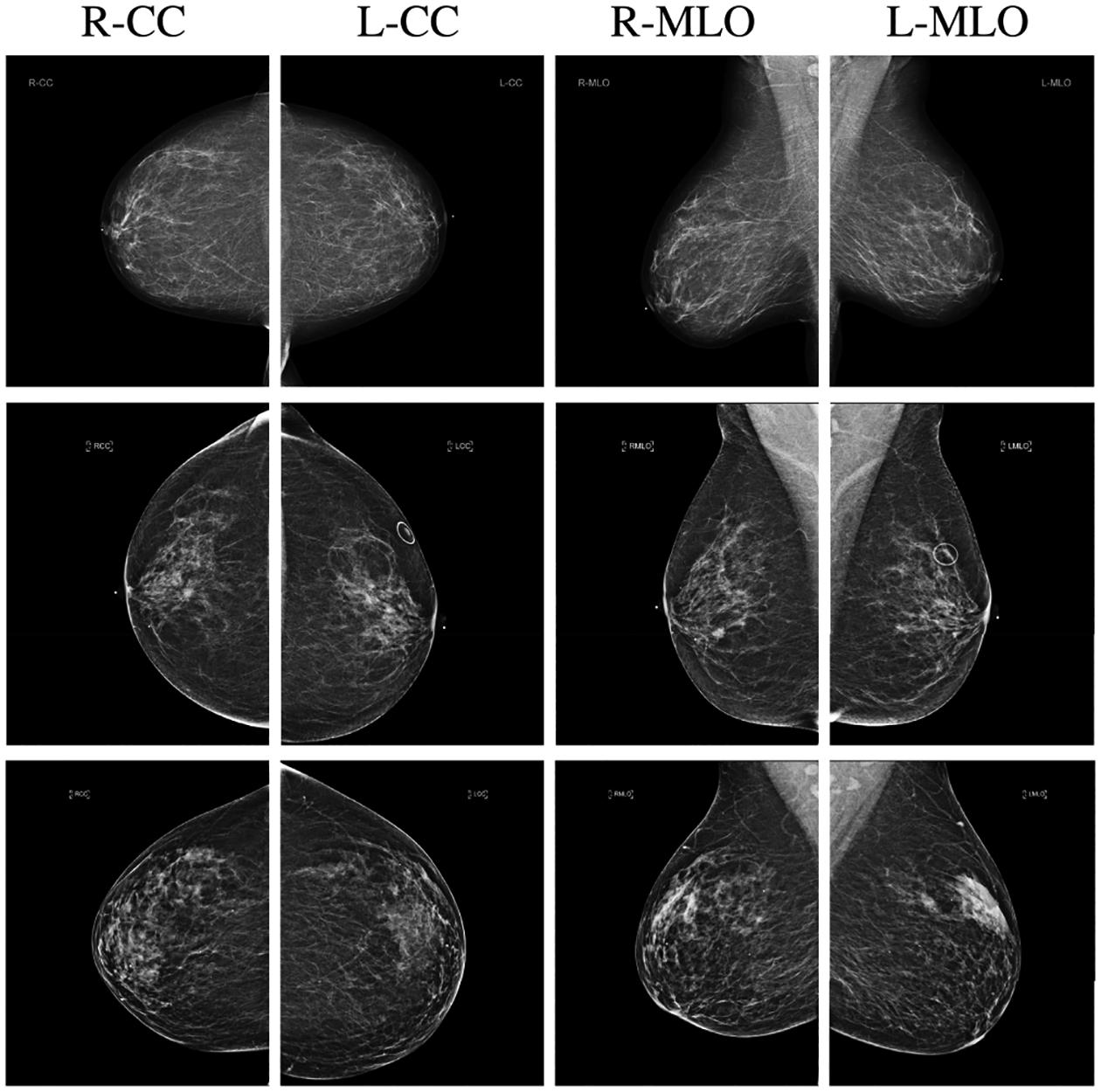
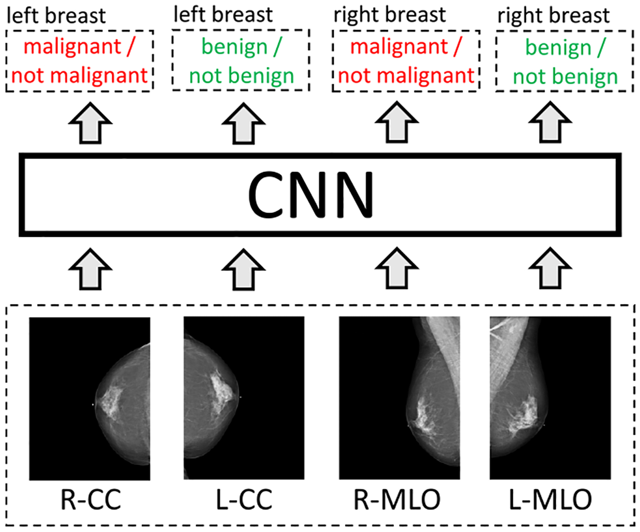
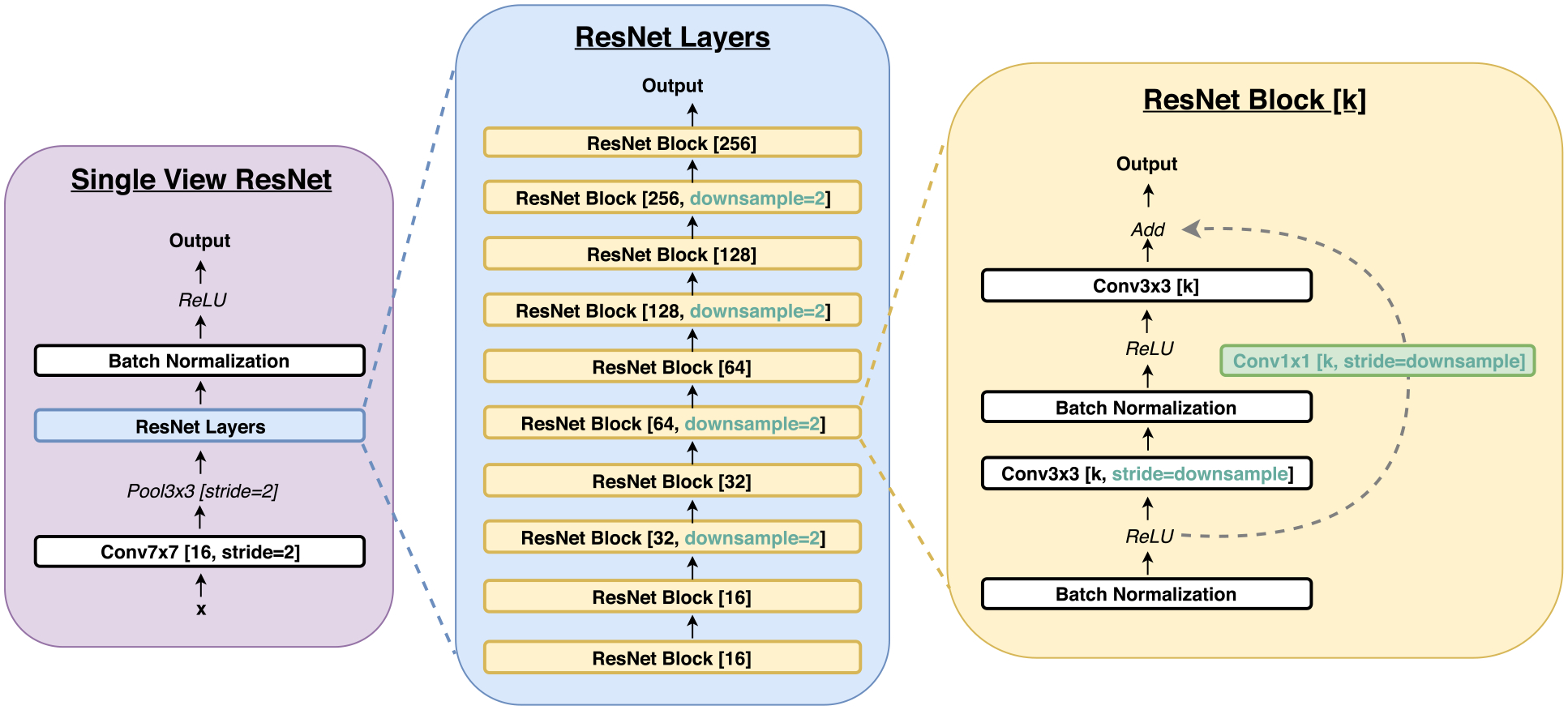
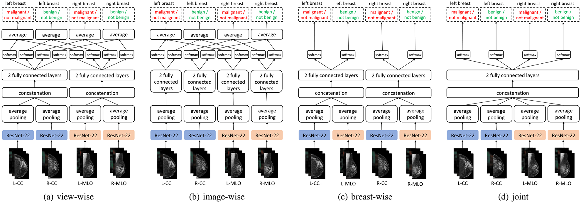
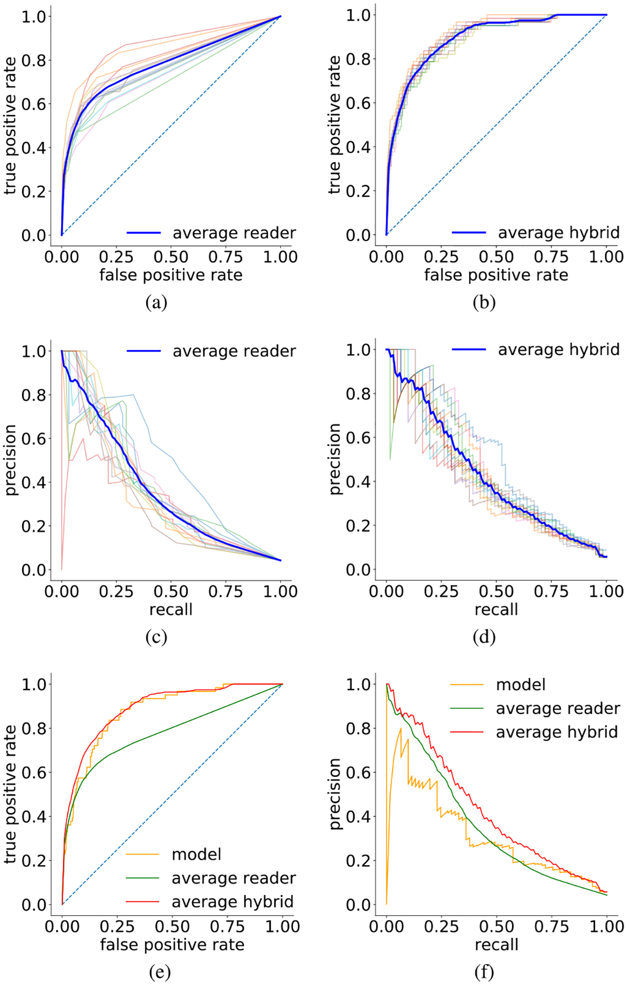
We present a deep convolutional neural network for breast cancer screening exam classification, trained, and evaluated on over 200000 exams (over 1000000 images). Our network achieves an AUC of 0.895 in predicting the presence of cancer in the breast, when tested on the screening population. We attribute the high accuracy to a few technical advances. 1) Our network's novel two-stage architecture and training procedure, which allows us to use a high-capacity patch-level network to learn from pixel-level labels alongside a network learning from macroscopic breast-level labels. 2) A custom ResNet-based network used as a building block of our model, whose balance of depth and width is optimized for high-resolution medical images. 3) Pretraining the network on screening BI-RADS classification, a related task with more noisy labels. 4) Combining multiple input views in an optimal way among a number of possible choices. To validate our model, we conducted a reader study with 14 readers, each reading 720 screening mammogram exams, and show that our model is as accurate as experienced radiologists when presented with the same data. We also show that a hybrid model, averaging the probability of malignancy predicted by a radiologist with a prediction of our neural network, is more accurate than either of the two separately. To further understand our results, we conduct a thorough analysis of our network's performance on different subpopulations of the screening population, the model's design, training procedure, errors, and properties of its internal representations. Our best models are publicly available at https://github.com/nyukat/breast_cancer_classifier.

摘要

我们提出了一种用于乳腺癌筛查检查分类的深度卷积神经网络,该网络在超过 200000 次检查(超过 1000000 张图像)上进行了训练和评估。当在筛查人群中进行测试时,我们的网络在预测乳腺癌的存在方面达到了 0.895 的 AUC。我们将高精度归因于一些技术进步。1)我们的网络新颖的两级架构和训练程序,允许我们使用大容量的补丁级网络从像素级标签和从宏观乳房级标签学习的网络学习。2)我们的模型中使用的自定义 ResNet 网络作为构建块,其深度和宽度的平衡针对高分辨率医学图像进行了优化。3)在筛查 BI-RADS 分类上对网络进行预训练,这是一项具有更多噪声标签的相关任务。4)在许多可能的选择中以最佳方式组合多个输入视图。为了验证我们的模型,我们进行了一项有 14 位读者参与的读者研究,每位读者阅读了 720 次筛查乳房 X 光检查,并表明当呈现相同数据时,我们的模型与经验丰富的放射科医生一样准确。我们还表明,将放射科医生预测的恶性肿瘤概率与我们的神经网络预测进行平均的混合模型比两者单独使用更准确。为了进一步了解我们的结果,我们对我们的网络在筛查人群的不同子群体、模型设计、训练程序、错误以及其内部表示的特性方面的性能进行了全面分析。我们的最佳模型可在 https://github.com/nyukat/breast_cancer_classifier 上获得。

https://cdn.ncbi.nlm.nih.gov/pmc/blobs/ca9f/7427471/eae5ec362025/nihms-1612554-f0010.jpg
https://cdn.ncbi.nlm.nih.gov/pmc/blobs/ca9f/7427471/0d15cabac1e8/nihms-1612554-f0001.jpg
https://cdn.ncbi.nlm.nih.gov/pmc/blobs/ca9f/7427471/12ed30afc167/nihms-1612554-f0002.jpg
https://cdn.ncbi.nlm.nih.gov/pmc/blobs/ca9f/7427471/60d756f8b29d/nihms-1612554-f0003.jpg
https://cdn.ncbi.nlm.nih.gov/pmc/blobs/ca9f/7427471/b60a2c1475cb/nihms-1612554-f0004.jpg
https://cdn.ncbi.nlm.nih.gov/pmc/blobs/ca9f/7427471/36b682005bb5/nihms-1612554-f0005.jpg
https://cdn.ncbi.nlm.nih.gov/pmc/blobs/ca9f/7427471/73a8b8e4e1b5/nihms-1612554-f0006.jpg
https://cdn.ncbi.nlm.nih.gov/pmc/blobs/ca9f/7427471/53ef2a4fc9b2/nihms-1612554-f0007.jpg
https://cdn.ncbi.nlm.nih.gov/pmc/blobs/ca9f/7427471/52c1c44a69c1/nihms-1612554-f0008.jpg
https://cdn.ncbi.nlm.nih.gov/pmc/blobs/ca9f/7427471/6e8f00712fe9/nihms-1612554-f0009.jpg
https://cdn.ncbi.nlm.nih.gov/pmc/blobs/ca9f/7427471/eae5ec362025/nihms-1612554-f0010.jpg

相似文献

1
Deep Neural Networks Improve Radiologists' Performance in Breast Cancer Screening.深度神经网络可提高放射科医生在乳腺癌筛查中的表现。
IEEE Trans Med Imaging. 2020 Apr;39(4):1184-1194. doi: 10.1109/TMI.2019.2945514. Epub 2019 Oct 7.
2
Understanding Clinical Mammographic Breast Density Assessment: a Deep Learning Perspective.理解临床乳腺 X 线摄影乳房密度评估:深度学习视角。
J Digit Imaging. 2018 Aug;31(4):387-392. doi: 10.1007/s10278-017-0022-2.
3
Can a Machine Learn from Radiologists' Visual Search Behaviour and Their Interpretation of Mammograms-a Deep-Learning Study.机器能否从放射科医生的视觉搜索行为及其对乳房 X 光照片的解释中学习——一项深度学习研究。
J Digit Imaging. 2019 Oct;32(5):746-760. doi: 10.1007/s10278-018-00174-z.
4
A deep learning method for classifying mammographic breast density categories.一种用于对乳腺钼靶图像的乳房密度类别进行分类的深度学习方法。
Med Phys. 2018 Jan;45(1):314-321. doi: 10.1002/mp.12683. Epub 2017 Dec 22.
5
Evaluation of Combined Artificial Intelligence and Radiologist Assessment to Interpret Screening Mammograms.联合人工智能和放射科医生评估解读筛查性乳房 X 光照片的效果。
JAMA Netw Open. 2020 Mar 2;3(3):e200265. doi: 10.1001/jamanetworkopen.2020.0265.
6
Comprehensive Word-Level Classification of Screening Mammography Reports Using a Neural Network Sequence Labeling Approach.基于神经网络序列标注方法的乳腺 X 线摄影筛查报告的全面词级分类。
J Digit Imaging. 2019 Oct;32(5):685-692. doi: 10.1007/s10278-018-0141-4.
7
A deep learning framework to classify breast density with noisy labels regularization.一种使用带噪标签正则化的深度学习框架来分类乳腺密度。
Comput Methods Programs Biomed. 2022 Jun;221:106885. doi: 10.1016/j.cmpb.2022.106885. Epub 2022 May 12.
8
Discriminating solitary cysts from soft tissue lesions in mammography using a pretrained deep convolutional neural network.使用预训练的深度卷积神经网络在乳腺钼靶摄影中区分孤立性囊肿与软组织病变。
Med Phys. 2017 Mar;44(3):1017-1027. doi: 10.1002/mp.12110.
9
A High-Performance Deep Neural Network Model for BI-RADS Classification of Screening Mammography.一种用于乳腺 X 线筛查 BI-RADS 分类的高性能深度神经网络模型。
Sensors (Basel). 2022 Feb 3;22(3):1160. doi: 10.3390/s22031160.
10
Can we reduce the workload of mammographic screening by automatic identification of normal exams with artificial intelligence? A feasibility study.能否通过人工智能自动识别正常的乳腺 X 光检查来减少工作量?一项可行性研究。
Eur Radiol. 2019 Sep;29(9):4825-4832. doi: 10.1007/s00330-019-06186-9. Epub 2019 Apr 16.

引用本文的文献

1
Deep Learning for the Early Detection of Invasive Ductal Carcinoma in Histopathological Images: Convolutional Neural Network Approach With Transfer Learning.基于深度学习的组织病理学图像中浸润性导管癌早期检测:采用迁移学习的卷积神经网络方法
JMIR Form Res. 2025 Aug 21;9:e62996. doi: 10.2196/62996.
2
Combined use of two artificial intelligence-based algorithms for mammography triaging: a retrospective simulation study.两种基于人工智能的算法联合用于乳腺钼靶筛查分诊:一项回顾性模拟研究。
Eur Radiol. 2025 Aug 21. doi: 10.1007/s00330-025-11849-x.
3
Exploring Gynecological Pathology and Imaging: A Crossroads of Technology, Biology, and Care.

本文引用的文献

1
Stand-Alone Artificial Intelligence for Breast Cancer Detection in Mammography: Comparison With 101 Radiologists.孤立人工智能在乳腺钼靶摄影中的乳腺癌检测:与 101 位放射科医生的比较。
J Natl Cancer Inst. 2019 Sep 1;111(9):916-922. doi: 10.1093/jnci/djy222.
2
New Frontiers: An Update on Computer-Aided Diagnosis for Breast Imaging in the Age of Artificial Intelligence.新前沿:人工智能时代下乳腺成像计算机辅助诊断的最新进展。
AJR Am J Roentgenol. 2019 Feb;212(2):300-307. doi: 10.2214/AJR.18.20392.
3
Detection of Breast Cancer with Mammography: Effect of an Artificial Intelligence Support System.
探索妇科病理学与影像学:技术、生物学与护理的交叉点
Diagnostics (Basel). 2025 Aug 7;15(15):1976. doi: 10.3390/diagnostics15151976.
4
ES-UNet: efficient 3D medical image segmentation with enhanced skip connections in 3D UNet.ES-UNet:通过在3D UNet中增强跳跃连接实现高效的3D医学图像分割
BMC Med Imaging. 2025 Aug 13;25(1):327. doi: 10.1186/s12880-025-01857-0.
5
Cancer Detection in Breast MRI Screening via Explainable AI Anomaly Detection.通过可解释人工智能异常检测在乳腺MRI筛查中进行癌症检测。
Radiology. 2025 Jul;316(1):e241629. doi: 10.1148/radiol.241629.
6
An Exploration of Discrepant Recalls Between AI and Human Readers of Malignant Lesions in Digital Mammography Screening.数字化乳腺钼靶筛查中人工智能与人类读者对恶性病变的差异召回率探究。
Diagnostics (Basel). 2025 Jun 19;15(12):1566. doi: 10.3390/diagnostics15121566.
7
AI in 2D Mammography: Improving Breast Cancer Screening Accuracy.二维乳腺钼靶摄影中的人工智能:提高乳腺癌筛查准确性
Medicina (Kaunas). 2025 Apr 26;61(5):809. doi: 10.3390/medicina61050809.
8
Bayesian-Optimized Convolutional Neural Networks for Classifying Primary Tumor Origin of Brain Metastases from MRI.用于通过磁共振成像(MRI)对脑转移瘤原发肿瘤起源进行分类的贝叶斯优化卷积神经网络
Brain Sci. 2025 Apr 25;15(5):450. doi: 10.3390/brainsci15050450.
9
Comparative Evaluation of Machine Learning-Based Radiomics and Deep Learning for Breast Lesion Classification in Mammography.基于机器学习的放射组学和深度学习在乳腺钼靶摄影中对乳腺病变分类的比较评估
Diagnostics (Basel). 2025 Apr 9;15(8):953. doi: 10.3390/diagnostics15080953.
10
Local Extremum Mapping for Weak Supervision Learning on Mammogram Classification and Localization.用于乳房X光片分类与定位的弱监督学习的局部极值映射
Bioengineering (Basel). 2025 Mar 21;12(4):325. doi: 10.3390/bioengineering12040325.
乳腺 X 线摄影术检测乳腺癌:人工智能支持系统的效果。
Radiology. 2019 Feb;290(2):305-314. doi: 10.1148/radiol.2018181371. Epub 2018 Nov 20.
4
Detecting and classifying lesions in mammograms with Deep Learning.深度学习在乳腺 X 光片中检测和分类病灶。
Sci Rep. 2018 Mar 15;8(1):4165. doi: 10.1038/s41598-018-22437-z.
5
Applying Data-driven Imaging Biomarker in Mammography for Breast Cancer Screening: Preliminary Study.应用数据驱动的乳腺成像生物标志物进行乳腺癌筛查:初步研究。
Sci Rep. 2018 Feb 9;8(1):2762. doi: 10.1038/s41598-018-21215-1.
6
Classifying symmetrical differences and temporal change for the detection of malignant masses in mammography using deep neural networks.使用深度神经网络对乳腺X线摄影中恶性肿块检测的对称差异和时间变化进行分类。
J Med Imaging (Bellingham). 2017 Oct;4(4):044501. doi: 10.1117/1.JMI.4.4.044501. Epub 2017 Oct 10.
7
Malignancy Detection on Mammography Using Dual Deep Convolutional Neural Networks and Genetically Discovered False Color Input Enhancement.使用双深度卷积神经网络和基因发现的假彩色输入增强技术进行乳腺钼靶摄影中的恶性肿瘤检测。
J Digit Imaging. 2017 Aug;30(4):499-505. doi: 10.1007/s10278-017-9993-2.
8
Deep Learning in Mammography: Diagnostic Accuracy of a Multipurpose Image Analysis Software in the Detection of Breast Cancer.深度学习在乳腺 X 线摄影中的应用:一种多功能图像分析软件在乳腺癌检测中的诊断准确性。
Invest Radiol. 2017 Jul;52(7):434-440. doi: 10.1097/RLI.0000000000000358.
9
Large scale deep learning for computer aided detection of mammographic lesions.基于大规模深度学习的计算机辅助乳腺病变检测
Med Image Anal. 2017 Jan;35:303-312. doi: 10.1016/j.media.2016.07.007. Epub 2016 Aug 2.
10
Diagnostic Accuracy of Digital Screening Mammography With and Without Computer-Aided Detection.有和没有计算机辅助检测的数字化乳腺筛查钼靶摄影的诊断准确性
JAMA Intern Med. 2015 Nov;175(11):1828-37. doi: 10.1001/jamainternmed.2015.5231.