Department of Pathology, University of Texas Medical Branch, Galveston, Texas, United States of America.
Department of Microbiology and Immunology, University of Texas Medical Branch, Galveston, Texas, United States of America.
PLoS Negl Trop Dis. 2020 Mar 2;14(3):e0007675. doi: 10.1371/journal.pntd.0007675. eCollection 2020 Mar.
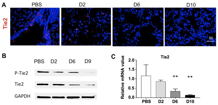
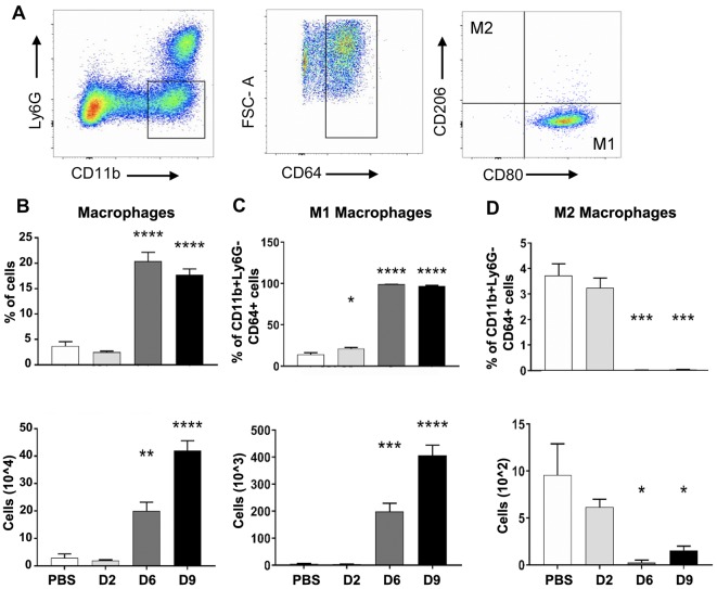
Orientia tsutsugamushi infection can cause acute lung injury and high mortality in humans; however, the underlying mechanisms are unclear. Here, we tested a hypothesis that dysregulated pulmonary inflammation and Tie2-mediated endothelial malfunction contribute to lung damage. Using a murine model of lethal O. tsutsugamushi infection, we demonstrated pathological characteristics of vascular activation and tissue damage: 1) a significant increase of ICAM-1 and angiopoietin-2 (Ang2) proteins in inflamed tissues and lung-derived endothelial cells (EC), 2) a progressive loss of endothelial quiescent and junction proteins (Ang1, VE-cadherin/CD144, occuludin), and 3) a profound impairment of Tie2 receptor at the transcriptional and functional levels. In vitro infection of primary human EC cultures and serum Ang2 proteins in scrub typhus patients support our animal studies, implying endothelial dysfunction in severe scrub typhus. Flow cytometric analyses of lung-recovered cells further revealed that pulmonary macrophages (MΦ) were polarized toward an M1-like phenotype (CD80+CD64+CD11b+Ly6G-) during the onset of disease and prior to host death, which correlated with the significant loss of CD31+CD45- ECs and M2-like (CD206+CD64+CD11b+Ly6G-) cells. In vitro studies indicated extensive bacterial replication in M2-type, but not M1-type, MΦs, implying the protective and pathogenic roles of M1-skewed responses. This is the first detailed investigation of lung cellular immune responses during acute O. tsutsugamushi infection. It uncovers specific biomarkers for vascular dysfunction and M1-skewed inflammatory responses, highlighting future therapeutic research for the control of this neglected tropical disease.
恙虫病东方体感染可导致人类急性肺损伤和高死亡率;然而,其潜在机制尚不清楚。在这里,我们验证了一个假设,即失调的肺部炎症和 Tie2 介导的内皮功能障碍导致肺部损伤。我们使用致死性恙虫病东方体感染的小鼠模型,证明了血管激活和组织损伤的病理特征:1)在炎症组织和肺源性内皮细胞(EC)中,ICAM-1 和血管生成素-2(Ang2)蛋白显著增加;2)内皮静止和连接蛋白(Ang1、VE-钙粘蛋白/CD144、occludin)逐渐丢失;3)Tie2 受体在转录和功能水平上受到严重损害。对原代人 EC 培养物的体外感染和恙虫病患者的血清 Ang2 蛋白支持了我们的动物研究,表明严重恙虫病存在内皮功能障碍。对肺回收细胞的流式细胞分析进一步表明,在疾病发作和宿主死亡之前,肺巨噬细胞(MΦ)向 M1 样表型(CD80+CD64+CD11b+Ly6G-)极化,这与 CD31+CD45-ECs 和 M2 样(CD206+CD64+CD11b+Ly6G-)细胞的显著减少相关。体外研究表明,M2 型而不是 M1 型 MΦ中存在广泛的细菌复制,这暗示了 M1 偏向反应的保护和致病作用。这是首次对急性恙虫病东方体感染期间肺部细胞免疫反应的详细研究。它揭示了血管功能障碍和 M1 偏向炎症反应的特定生物标志物,突出了控制这种被忽视的热带病的未来治疗研究。