Radiology and Diagnostic Imaging Department, IRCCS Regina Elena National Cancer Institute, Rome, Italy.
Departmental Faculty of Medicine and Surgery, Center for Integrated Research, University Campus Bio-Medico of Rome, Rome, Italy.
PLoS One. 2020 Mar 2;15(3):e0229611. doi: 10.1371/journal.pone.0229611. eCollection 2020.
To investigate the correlation between histogram-based Dynamic Contrast-Enhanced magnetic resonance imaging (DCE-MRI) parameters and positron emission tomography with 18F-fluorodeoxyglucose (18F-FDG-PET) values in oropharyngeal squamous cell carcinoma (OPSCC), both in primary tumors (PTs) and in metastatic lymph nodes (LNs).
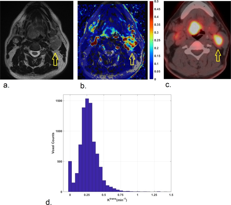
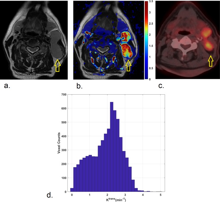
52 patients with a new pathologically-confirmed OPSCC were included in the present retrospective cohort study. Imaging including DCE-MRI and 18F-FDG PET/CT scans were acquired in all patients. Both PTs and the largest LN, if present, were volumetrically contoured. Quantitative parameters, including the transfer constants, Ktrans and Kep, and the volume of extravascular extracellular space, ve, were calculated from DCE-MRI. The percentiles (P), P10, P25, P50, P75, P90, and skewness, kurtosis and entropy were obtained from the histogram-based analysis of each perfusion parameter. Standardized uptake values (SUV), SUVmax, SUVpeak, SUVmean, metabolic tumor volume (MTV) and total lesion glycolysis (TLG) were calculated applying a SUV threshold of 40%. The correlations between all variables were investigated with the Spearman-rank correlation test. To exclude false positive results under multiple testing, the Benjamini-Hockberg procedure was applied.
No significant correlations were found between any parameters in PTs, while significant associations emerged between Ktrans and 18F-FDG PET parameters in LNs.
Evident relationships emerged between DCE-MRI and 18F-FDG PET parameters in OPSCC LNs, while no association was found in PTs. The complex relationships between perfusion and metabolic biomarkers should be interpreted separately for primary tumors and lymph-nodes. A multiparametric approach to analyze PTs and LNs before treatment is advisable in head and neck squamous cell carcinoma (HNSCC).
探讨头颈部鳞癌(HNSCC)原发灶(PT)和转移性淋巴结(LN)中动态对比增强磁共振成像(DCE-MRI)基于直方图的参数与正电子发射断层扫描(PET)18F-氟代脱氧葡萄糖(18F-FDG-PET)值之间的相关性。
本回顾性队列研究共纳入 52 例经病理证实的新发性口咽鳞状细胞癌患者。所有患者均行 DCE-MRI 和 18F-FDG PET/CT 扫描。对所有肿瘤进行容积勾画,包括 PT 和最大的 LN。从 DCE-MRI 计算获得转移常数 Ktrans 和 Kep 以及血管外细胞外容积 ve 等定量参数。通过各灌注参数的直方图分析获得百分位数(P)、P10、P25、P50、P75、P90 和偏度、峰度和熵。应用 SUV 阈值为 40%计算标准化摄取值(SUV)、SUVmax、SUVpeak、SUVmean、代谢肿瘤体积(MTV)和总病变糖酵解(TLG)。采用 Spearman 秩相关检验分析所有变量之间的相关性。为排除多重检验中的假阳性结果,采用 Benjamini-Hockberg 程序。
PT 中各参数之间无显著相关性,而 LN 中 Ktrans 与 18F-FDG PET 参数之间存在显著相关性。
OPSCC LN 中 DCE-MRI 与 18F-FDG PET 参数之间存在明显关系,而在 PT 中未发现这种关系。在分析头颈部鳞癌的 PT 和 LN 时,灌注和代谢生物标志物之间的复杂关系应分别进行解释。在治疗前对头颈部鳞癌的 PT 和 LN 进行多参数分析是明智的。