Multimodality Medical Imaging M3i Group, Faculty of Science and Technology, Technical Medical Centre, University of Twente, PO Box 217, Enschede, The Netherlands.
Robotics and Mechatronics Group, Faculty of Electrical Engineering, Mathematics, and Computer Science, Technical Medical Centre, University of Twente, Enschede, The Netherlands.
Eur Radiol Exp. 2020 Mar 4;4(1):15. doi: 10.1186/s41747-019-0133-2.
We aimed at reviewing design and realisation of perfusion/flow phantoms for validating quantitative perfusion imaging (PI) applications to encourage best practices.
A systematic search was performed on the Scopus database for "perfusion", "flow", and "phantom", limited to articles written in English published between January 1999 and December 2018. Information on phantom design, used PI and phantom applications was extracted.
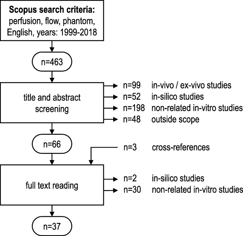
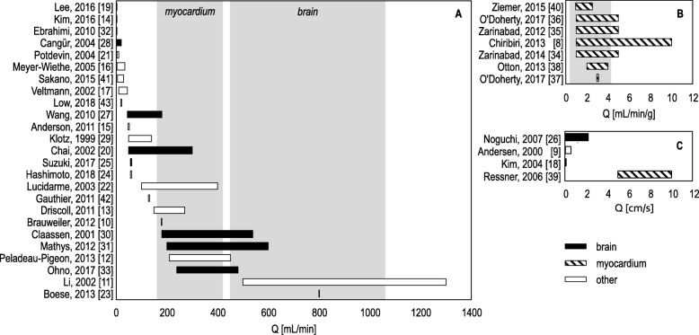
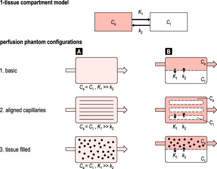
Of 463 retrieved articles, 397 were rejected after abstract screening and 32 after full-text reading. The 37 accepted articles resulted to address PI simulation in brain (n = 11), myocardial (n = 8), liver (n = 2), tumour (n = 1), finger (n = 1), and non-specific tissue (n = 14), with diverse modalities: ultrasound (n = 11), computed tomography (n = 11), magnetic resonance imaging (n = 17), and positron emission tomography (n = 2). Three phantom designs were described: basic (n = 6), aligned capillary (n = 22), and tissue-filled (n = 12). Microvasculature and tissue perfusion were combined in one compartment (n = 23) or in two separated compartments (n = 17). With the only exception of one study, inter-compartmental fluid exchange could not be controlled. Nine studies compared phantom results with human or animal perfusion data. Only one commercially available perfusion phantom was identified.
We provided insights into contemporary phantom approaches to PI, which can be used for ground truth evaluation of quantitative PI applications. Investigators are recommended to verify and validate whether assumptions underlying PI phantom modelling are justified for their intended phantom application.
本研究旨在回顾用于验证定量灌注成像(PI)应用的灌注/流动体模的设计和实现,以鼓励最佳实践。
我们在 Scopus 数据库中针对“灌注”、“流动”和“体模”进行了系统检索,检索范围限于 1999 年 1 月至 2018 年 12 月期间以英文发表的文章。提取了体模设计、使用的 PI 和体模应用的信息。
在检索到的 463 篇文章中,经过摘要筛选后有 397 篇被排除,全文阅读后又有 32 篇被排除。最终纳入的 37 篇文章涉及 PI 在脑(n = 11)、心肌(n = 8)、肝(n = 2)、肿瘤(n = 1)、手指(n = 1)和非特定组织(n = 14)中的模拟,应用的模态有超声(n = 11)、计算机断层扫描(n = 11)、磁共振成像(n = 17)和正电子发射断层扫描(n = 2)。描述了三种体模设计:基本型(n = 6)、对准毛细管型(n = 22)和组织填充型(n = 12)。微血管和组织灌注在一个腔室中(n = 23)或在两个分开的腔室中(n = 17)组合。除了一项研究外,腔室间的液体交换均无法控制。有 9 项研究比较了体模结果与人体或动物的灌注数据。只发现了一种市售的灌注体模。
我们深入了解了 PI 当代体模方法,可以用于定量 PI 应用的真实数据评估。建议研究人员验证和确认 PI 体模建模的假设是否适用于其预期的体模应用。